. 2015 Mar 15;308(6):F594-601.
doi: 10.1152/ajprenal.00609.2014. Epub 2015 Jan 7.

An angiotensin-(1-7) peptidase in the kidney cortex, proximal tubules, and human HK-2 epithelial cells that is distinct from insulin-degrading enzyme

Affiliations

An angiotensin-(1-7) peptidase in the kidney cortex, proximal tubules, and human HK-2 epithelial cells that is distinct from insulin-degrading enzyme

Bryan A Wilson et al. Am J Physiol Renal Physiol. .

Abstract

Angiotensin 1-7 [ANG-(1-7)] is expressed within the kidney and exhibits renoprotective actions that antagonize the inflammatory, fibrotic, and pro-oxidant effects of ANG II. We previously identified an peptidase that preferentially metabolized ANG-(1-7) to ANG-(1-4) in the brain medulla and cerebrospinal fluid (CSF) of sheep (Marshall AC, Pirro NT, Rose JC, Diz DI, Chappell MC. J Neurochem 130: 313-323, 2014); thus the present study established the expression of the peptidase in the kidney. Utilizing a sensitive HPLC-based approach, we demonstrate a peptidase activity that hydrolyzed ANG-(1-7) to ANG-(1-4) in the sheep cortex, isolated tubules, and human HK-2 renal epithelial cells. The peptidase was markedly sensitive to the metallopeptidase inhibitor JMV-390; human HK-2 cells expressed subnanomolar sensitivity (IC50 = 0.5 nM) and the highest specific activity (123 ± 5 fmol·min(-1)·mg(-1)) compared with the tubules (96 ± 12 fmol·min(-1)·mg(-1)) and cortex (107 ± 9 fmol·min(-1)·mg(-1)). The peptidase was purified 41-fold from HK-2 cells; the activity was sensitive to JMV-390, the chelator o-phenanthroline, and the mercury-containing compound p-chloromercuribenzoic acid (PCMB), but not to selective inhibitors against neprilysin, neurolysin and thimet oligopeptidase. Both ANG-(1-7) and its endogenous analog [Ala(1)]-ANG-(1-7) (alamandine) were preferentially hydrolyzed by the peptidase compared with ANG II, [Asp(1)]-ANG II, ANG I, and ANG-(1-12). Although the ANG-(1-7) peptidase and insulin-degrading enzyme (IDE) share similar inhibitor characteristics of a metallothiolendopeptidase, we demonstrate marked differences in substrate specificity, which suggest these peptidases are distinct. We conclude that an ANG-(1-7) peptidase is expressed within the renal proximal tubule and may play a potential role in the renal renin-angiotensin system to regulate ANG-(1-7) tone.

Keywords: ANG-(1–7); HK-2 epithelial cells; endopeptidase; proximal tubules.


Figures

Fig. 1.
Fig. 1.
ANG-(1–7) peptidase activity in the sheep kidney cortex and isolated proximal tubules. 125I-ANG-(1–7) (125I-A7) was hydrolyzed to 125I-ANG-(1–4) (125I-A4) in the supernatant from kidney cortex (A) and was reduced by JMV-390 (1 μM; B); 125I-A7 was hydrolyzed to 125I-A4 in the supernatant from isolated proximal tubules (C) and was reduced by JMV-390 (1 μM; D). Peptidase assays were conducted in the presence of an inhibitor cocktail for 60 min at 37°C, and products were detected by HPLC-γ detector under isocratic conditions. Data are representative of 3 separate experiments from different animals (see Table 1).
Fig. 2.
Fig. 2.
ANG-(1–7) peptidase activity in human HK-2 cell supernatant and media. 125I-A7 was hydrolyzed to 125I-A4 and 125I-ANG-(3–4), the small peak eluting before 125I-A4 in the HK-2 100,000-g cytosol fraction (A) and was abolished by JMV-390 (1 μM; B). 125I-A7 was hydrolyzed to 125I-A4 in the media from HK-2 cells (C) and was abolished by JMV-390 (1 μM; D). Peptidase assays were conducted without additional inhibitors for 60 min at 37°C, and products were detected by HPLC-γ detector under isocratic conditions. Data are representative of 3 separate experiments from 3 distinct cell passages (see Table 1).
Fig. 3.
Fig. 3.
Characterization of ANG-(1–7) peptidase activity in human HK-2 cells. Dose-response curves for inhibition of 125I-ANG-(1–4) production by unlabeled ANG-(1–7) (A) and unlabeled [Ala1]-ANG-(1–7) (B) in HK-2 100,000-g cytosol fraction and dose-response curves for inhibition of 125I-ANG-(1–4) production by the JMV-390 inhibitor in HK-2 cell supernatant (C) and media (D) are shown. Values are means ± SE; n = 3. Monophasic curve fit and IC50 values were derived with GraphPad Prism 5.
Fig. 4.
Fig. 4.
Purified HK-2 peptidase preferentially hydrolyzes ANG-(1–7) to ANG-(1–4). Purified peptidase (0.3 ug) from the HK-2 100,000-g cytosol fraction was incubated with 100 μM of ANG-(1–7) (A7; A); A7+1 μM JMV-390 (B); [Ala1]-ANG-(1–7) (Ala1-A7; C); ANG II (AII; D); ANG I (AI; E); and ANG-(1–12) (A12; F). Reactions occurred for 20 h at 37°C, and the products were detected by HPLC-UV (220 nM) under identical gradient conditions.
Fig. 5.
Fig. 5.
Comparison of the peptidase velocities for the hydrolysis of angiotensin peptides by purified HK-2 peptidase. Purified peptidase (0.3 μg) was incubated with angiotensin peptides (100 μM), and metabolism was detected by HPLC-UV (220 nM) under gradient conditions. Data are from a single experiment.
Fig. 6.
Fig. 6.
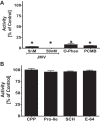
Inhibitor profile of purified HK-2 peptidase. Peptidase activity was blocked by JMV-390 (JMV, 5 and 50 nM) and the thiol inhibitor para-chloromercuribenzoic acetate (PCMB; 10 μM), and metallochelator ortho-phenanthroline (O-Phen; 10 μM; A). Other inhibitors including CPP (10 μM), Pro-Ile (10 μM), SCH (10 μM), and E-64 (10 μM) did not significantly inhibit activity (B). Peptidase reactions were performed with 125I-ANG-(1–7) for 60 min at 37°C. Values are means ± SE; n = 3. *P < 0.05 vs. control conditions.
Fig. 7.
Fig. 7.
ANG-(1–7) peptidase from HK-2 cells is distinct from insulin-degrading enzyme (IDE). Purified HK-2 peptidase (0.3 μg) hydrolyzed 125I-A7 to 125I-A4 (A) but did not hydrolyze Abz-ANG-(1–7)-[Tyr7 (NO2)] (Abz-A7; B). Human recombinant IDE (2 μg) did not hydrolyze 125I-A7 (C) but hydrolyzed Abz-A7 to Abz-ANG-(1–4) (Abz-A4; D). Products were separated by HPLC under isocratic (A and C) or gradient (B and D) conditions.
Fig. 8.
Fig. 8.
HK-2 processing of 125I-ANG I. Chromatograph reveals 125I-ANG I (125I-AI) is metabolized to 125I-A7 and 125I-A4 in the supernatant from HK-2 100,000-g cytosol fraction (A). 125I-A7 metabolism to 125I-A4 is reduced by 1 nM JMV-390 (B), and 125I-AI metabolism to 125I-A7 is reduced by 10 μM CPP in combination with 1 nM JMV-390 (C). AI metabolism is abolished by 10 μM PCMB (D). 125I-labeled products were separated by HPLC under gradient conditions.
Fig. 9.
Fig. 9.
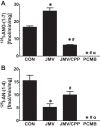
125I-ANG I metabolism in HK-2 cells. JMV-390 (JMV, 1 nM) increased 125I-A7, while CPP (10 μM) and JMV reduced 125I-A7 (A). JMV reduced 125I-A4 levels while addition of JMV and CPP did not further influence 125I-A4 (B). The mercury-containing inhibitor PCMB (10 μM) abolished the metabolism of 125I-ANG I to 125I-A7 and 125I-A4. Values are means ± SE; n = 3. *P < 0.05 vs. control (CON). #P < 0.05 vs. JMV. αP < 0.05 vs. JMV/CPP.

References

    1. Ali Q, Wu Y, Hussain T. Chronic AT2 receptor activation increases renal ACE2 activity, attenuates AT1 receptor function and blood pressure in obese Zucker rats. Kidney Int 84: 931–939, 2013. - PMC - PubMed
    1. Alzayadneh EM, Chappell MC. Angiotensin-(1–7) abolishes AGE-induced cellular hypertrophy and myofibroblast transformation via inhibition of ERK1/2. Cell Signal 26: 3027–3025, 2014. - PMC - PubMed
    1. Bader M. ACE2, angiotensin-(1–7), and Mas: the other side of the coin. Pflügers Arch 465: 79–85, 2013. - PubMed
    1. Bennett RG, Duckworth WC, Hamel FG. Degradation of amylin by insulin-degrading enzyme. J Biol Chem 275: 36621–36625, 2000. - PubMed
    1. Bennett RG, Heimann DG, Hamel FG. Degradation of relaxin family peptides by insulin-degrading enzyme. Ann NY Acad Sci 1160: 38–41, 2009. - PMC - PubMed

Publication types

LinkOut - more resources