. 2015 Sep;26(9):2277-88.
doi: 10.1681/ASN.2014070641. Epub 2015 Jan 7.

Podocyte Number in Children and Adults: Associations with Glomerular Size and Numbers of Other Glomerular Resident Cells

Affiliations

Podocyte Number in Children and Adults: Associations with Glomerular Size and Numbers of Other Glomerular Resident Cells

Victor G Puelles et al. J Am Soc Nephrol. 2015 Sep.

Abstract

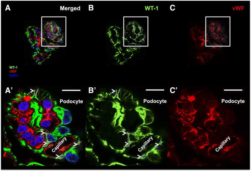
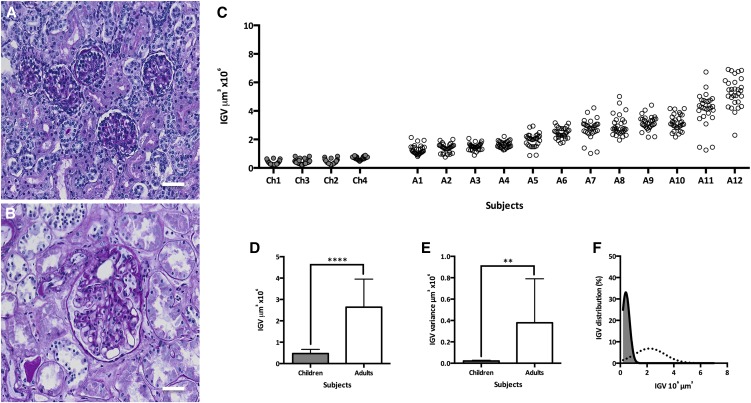
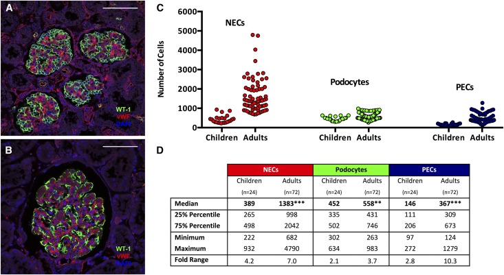
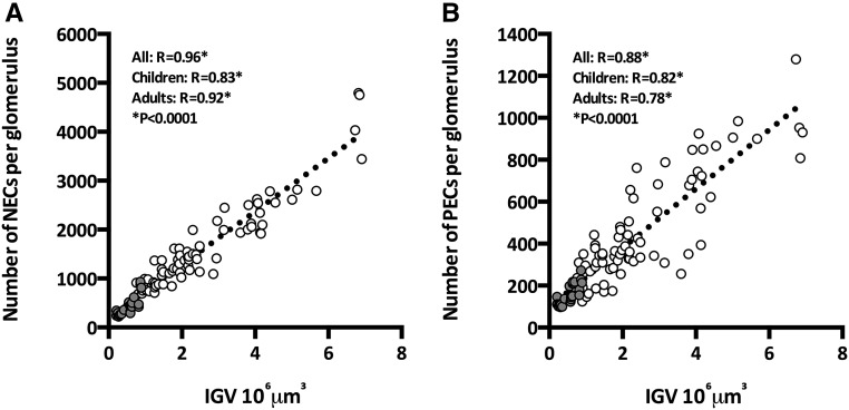
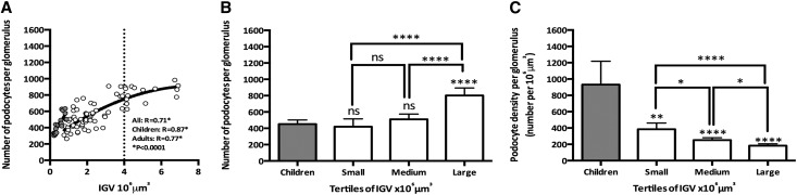
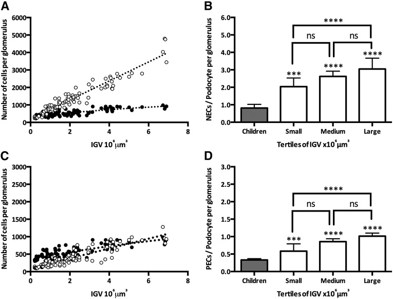
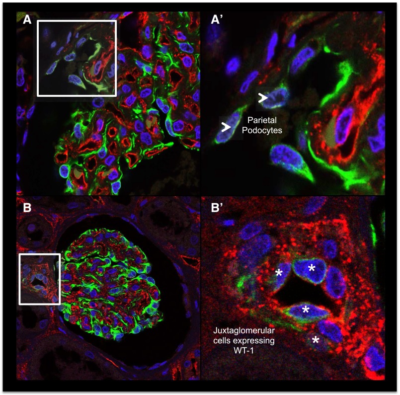
Increases in glomerular size occur with normal body growth and in many pathologic conditions. In this study, we determined associations between glomerular size and numbers of glomerular resident cells, with a particular focus on podocytes. Kidneys from 16 male Caucasian-Americans without overt renal disease, including 4 children (≤3 years old) to define baseline values of early life and 12 adults (≥18 years old), were collected at autopsy in Jackson, Mississippi. We used a combination of immunohistochemistry, confocal microscopy, and design-based stereology to estimate individual glomerular volume (IGV) and numbers of podocytes, nonepithelial cells (NECs; tuft cells other than podocytes), and parietal epithelial cells (PECs). Podocyte density was calculated. Data are reported as medians and interquartile ranges (IQRs). Glomeruli from children were small and contained 452 podocytes (IQR=335-502), 389 NECs (IQR=265-498), and 146 PECs (IQR=111-206). Adult glomeruli contained significantly more cells than glomeruli from children, including 558 podocytes (IQR=431-746; P<0.01), 1383 NECs (IQR=998-2042; P<0.001), and 367 PECs (IQR=309-673; P<0.001). However, large adult glomeruli showed markedly lower podocyte density (183 podocytes per 10(6) µm(3)) than small glomeruli from adults and children (932 podocytes per 10(6) µm(3); P<0.001). In conclusion, large adult glomeruli contained more podocytes than small glomeruli from children and adults, raising questions about the origin of these podocytes. The increased number of podocytes in large glomeruli does not match the increase in glomerular size observed in adults, resulting in relative podocyte depletion. This may render hypertrophic glomeruli susceptible to pathology.

Keywords: glomerular hyperfiltration; glomerulus; podocyte.


Figures

Figure 1.
Figure 1.
Podocyte identification. (A) A representative confocal image of a glomerular tuft is shown (merged) using (B) WT-1 (green; specific podocyte marker), (C) vWF (red; specific endothelial cell marker), and DAPI (blue; nuclear marker). The corresponding insets show our podocyte identification criteria: (A′) expression of WT-1 in podocyte cytoplasm, (B′) lack of expression of vWF, and (C′) their location outside capillaries. Arrowheads show classic podocyte morphology (major projections). Scale bars, 10 μm. DAPI, 4′,6-diamidino-2-phenylindole.
Figure 2.
Figure 2.
Glomeruli from adults are larger and have more variable size than glomeruli from children. A and B show representative images of average glomeruli from children and adults, respectively, using periodic acid–Schiff staining. (C) IGV values in 16 Caucasian-American men and boys. Each circle represents one glomerulus, and each column represents one subject with 30 glomeruli per subject; Ch1–Ch4 represent four young children (gray), and A1–A12 represent 12 adults (white) ranked by median IGV. (D) Aggregated IGV differences between children and adults. (E) Difference in IGV variance per subject between glomeruli from children and adults. (F) IGV distributions in children (solid line with gray area) and adults (dotted line). Bars represent median values with IQR. Scale bars, 50 μm. **P<0.01; ****P<0.001.
Figure 3.
Figure 3.
Large glomeruli from adults contained more podocytes, NECs and PECs than glomeruli from children. A and B show representative confocal images of glomeruli from children and adults, respectively, with WT-1 (green), vWF (red), and DAPI (blue). C and D show the numbers of NECs, PECs, and podocytes per glomerulus from children and adults. C illustrates the variability of each cell type between glomeruli from adults and children; each circle represents the value for a single glomerulus,. D provides details of the medians, IQRs (25% and 75% percentiles), minimum and maximum values, and fold ranges per group. Scale bars, 100 μm. DAPI, 4′,6-diamidino-2-phenylindole. **P<0.01; ***P<0.001.
Figure 4.
Figure 4.
Glomerular size is closely associated with numbers of NECs and PECs. (A) Association between IGV and numbers of NECs in glomeruli from children (gray) and adults (white). (B) Association between IGV and PECs in glomeruli from children (gray) and adults (white).
Figure 5.
Figure 5.
Large adult glomeruli have more podocytes and lower podocyte density than smaller adult glomeruli and glomeruli from children. (A) IGV and numbers of podocytes in children (gray circles) and adults (white circles). The solid line represents the line of best fit in children (linear) and adults (quadratic), and the dotted line shows the beginning of the curve plateau at 4×106 μm3. (B) Number of podocytes in children and adult IGV tertiles (small, medium, and large glomeruli). (C) Podocyte density in children and adult IGV tertiles (small, medium, and large glomeruli). Children: aggregated data from four infants (6 glomeruli per subject; n=24 glomeruli); adult IGV tertiles were tertile 1 (small glomeruli), between 0.76 and 1.72×106 μm3; tertile 2 (medium glomeruli), between 1.74 and 2.86×106 μm3; and tertile 3 (large glomeruli), between 2.94 and 6.91×106 μm3. P values directly over the bars represent comparisons between children (gray bars) and each adult tertile. Bars represent median values with IQR. *P<0.05; **P<0.01; ****P<0.001.
Figure 6.
Figure 6.
Numbers of NECs, PECs, and podocytes in the context of glomerular volume. (A) Numbers of podocytes (black circles) and NECs (white circles) in the context of IGV. (B) NEC-to-podocyte ratio in glomeruli from children and adults by adult IGV tertiles. (C) Numbers of PECs (black circles) and podocytes (white circles) in the context of IGV. (D) PEC-to-podocyte ratio in glomeruli from children and adults by adult IGV tertiles. P values directly over the bars represent comparisons between children (gray bars) and each adult tertile. Bars represent median values with IQR. ***P<0.001; ****P<0.001.
Figure 7.
Figure 7.
Possible podocyte sources in the human kidney. (A and A′) Parietal podocytes: PECs expressing WT-1 are located close to the vascular pole of a glomerulus (arrowheads). (B and B′) WT-1+cells were also occasionally observed in the tunica intima and the tunica media of arterioles in the JGA (asterisks).

References

    1. Saran R, Shahinian V: CKD: A pandemic calling for concerted public health action. Adv Chronic Kidney Dis 17: 213–214, 2010 - PubMed
    1. Wiggins RC: The spectrum of podocytopathies: A unifying view of glomerular diseases. Kidney Int 71: 1205–1214, 2007 - PubMed
    1. Reiser J, Sever S: Podocyte biology and pathogenesis of kidney disease. Annu Rev Med 64: 357–366, 2013 - PMC - PubMed
    1. Jefferson JA, Alpers CE, Shankland SJ: Podocyte biology for the bedside. Am J Kidney Dis 58: 835–845, 2011 - PMC - PubMed
    1. Wharram BL, Goyal M, Wiggins JE, Sanden SK, Hussain S, Filipiak WE, Saunders TL, Dysko RC, Kohno K, Holzman LB, Wiggins RC: Podocyte depletion causes glomerulosclerosis: Diphtheria toxin-induced podocyte depletion in rats expressing human diphtheria toxin receptor transgene. J Am Soc Nephrol 16: 2941–2952, 2005 - PubMed

Publication types

LinkOut - more resources