. 2015 Mar;89(6):3209-20.
doi: 10.1128/JVI.03072-14. Epub 2015 Jan 7.

Impact of the adenoviral E4 Orf3 protein on the activity and posttranslational modification of p53

Affiliations

Impact of the adenoviral E4 Orf3 protein on the activity and posttranslational modification of p53

Caroline J DeHart et al. J Virol. 2015 Mar.

Abstract

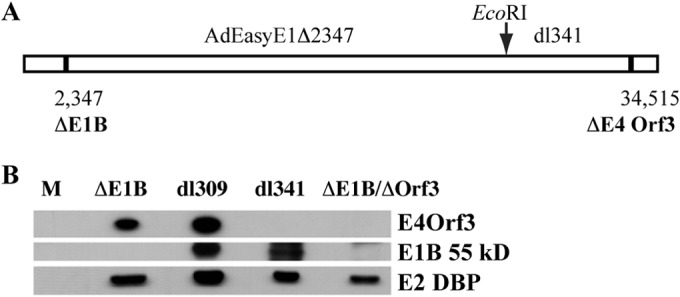
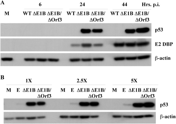
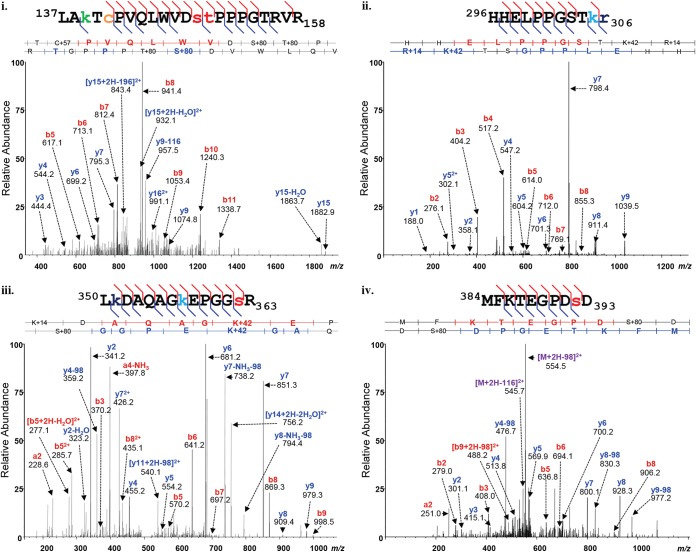
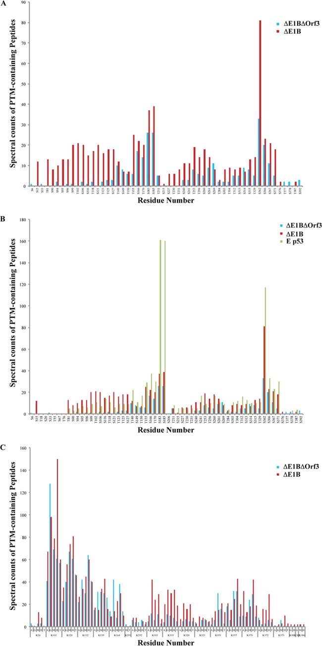
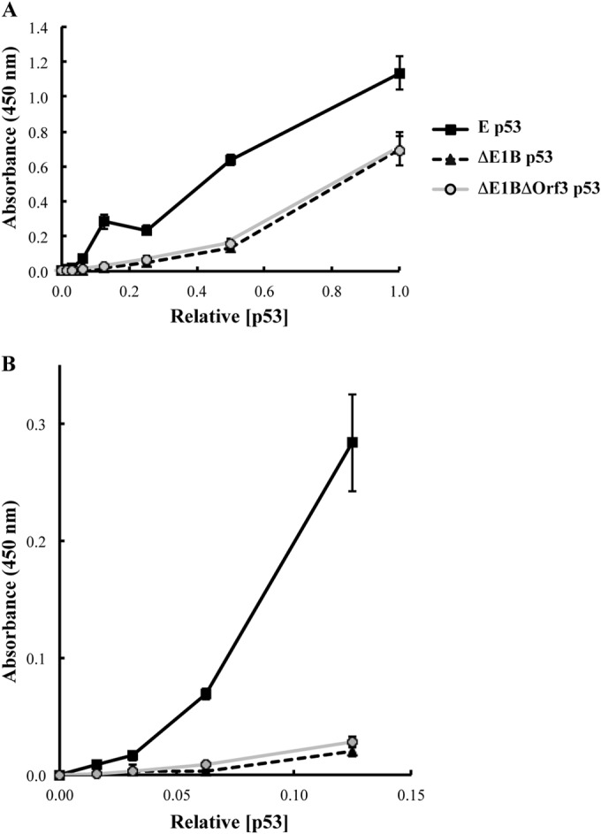
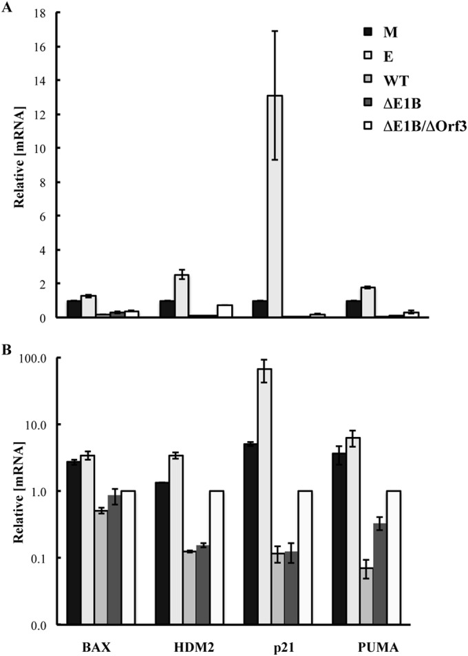
Our previous studies have established that the p53 populations that accumulate in normal human cells exposed to etoposide or infected by an E1B 55-kDa protein-null mutant of human adenovirus type 5 carry a large number of posttranslational modifications at numerous residues (C. J. DeHart, J. S. Chahal, S. J. Flint, and D. H. Perlman, Mol Cell Proteomics 13:1-17, 2014, http://dx.doi.org/10.1074/mcp.M113.030254). In the absence of this E1B protein, the p53 transcriptional program is not induced, and it has been reported that the viral E4 Orf3 protein inactivates p53 (C. Soria, F. E. Estermann, K. C. Espantman, and C. C. O'Shea, Nature 466:1076-1081, 2010, http://dx.doi.org/10.1038/nature09307). As the latter protein disrupts nuclear Pml bodies, sites at which p53 is modified, we used mass spectrometry to catalogue the posttranscriptional modifications of the p53 population that accumulates when neither the E1B 55-kDa nor the E4 Orf3 protein is made in infected cells. Eighty-five residues carrying 163 modifications were identified. The overall patterns of posttranslational modification of this population and p53 present in cells infected by an E1B 55-kDa-null mutant were similar. The efficiencies with which the two forms of p53 bound to a consensus DNA recognition sequence could not be distinguished and were lower than that of transcriptionally active p53. The absence of the E4 Orf3 protein increased expression of several p53-responsive genes when the E1B protein was also absent from infected cells. However, expression of these genes did not attain the levels observed when p53 was activated in response to etoposide treatment and remained lower than those measured in mock-infected cells.

Importance: The tumor suppressor p53, a master regulator of cellular responses to stress, is inactivated and destroyed in cells infected by species C human adenoviruses, such as type 5. It is targeted for proteasomal degradation by the action of a virus-specific E3 ubiquitin ligase that contains the viral E1B 55-kDa and E4 Orf6 proteins, while the E4 Orf3 protein has been reported to block its ability to stimulate expression of p53-dependent genes. The comparisons reported here of the posttranslational modifications and activities of p53 populations that accumulate in infected normal human cells in the absence of both mechanisms of inactivation or of only the E3 ligase revealed little impact of the E4 Orf3 protein. These observations indicate that E4 Orf3-dependent disruption of Pml bodies does not have a major effect on the pattern of p53 posttranslational modifications in adenovirus-infected cells. Furthermore, they suggest that one or more additional viral proteins contribute to blocking p53 activation and the consequences that are deleterious for viral reproduction, such as apoptosis or cell cycle arrest.


Figures

FIG 1
FIG 1
Construction and characterization of the E1B 55-kDa- and E4 Orf3-null double-mutant virus AdEasyE1Δ2347-dl341. (A) The origins of the segments of the double mutant virus genome are shown schematically, indicating the positions of the single-pair deletions that prevent production of the E1B 55-kDa or E4 Orf3 protein (ΔE1B and ΔE4 Orf3, respectively) and the EcoRI site exploited in the construction of the mutant. (B) HFFs were infected with 100 PFU/cell AdEasyE1Δ2347 (ΔE1B), dl309, dl341, or the double mutant (ΔE1B/ΔOrf3) for 24 h, and the viral proteins listed on the right were examined by immunoblotting.
FIG 2
FIG 2
Accumulation of p53 in AdEasyE1Δ2347-dl341-infected cells. (A) HFFs were infected with 100 PFU/cell wild-type AdEasyE1 (WT), AdEasyE1Δ2347 (ΔE1B), or AdEasyE1Δ2347-dl341 (ΔE1B/ΔOrf3) for the periods indicated or mock infected (M). Whole-cell lysates were prepared, and the proteins listed on the right were examined by immunoblotting. (B) HFFs were infected for 44 h with the viruses indicated, mock infected, or incubated with medium containing 125 μM etoposide (E) for 44 h. Cell lysates were prepared, and p53 present in the increasing lysate volumes indicated at the top was visualized by immunoblotting.
FIG 3
FIG 3
MS-MS spectra of p53 isolated from AdEasyE1Δ2347-dl341-infected cells. The p53 protein was isolated from double-mutant-infected cells and subjected to protease digestion as described in Materials and Methods. The peptides were analyzed using reversed-phase nano-UPLC-MS and MS-MS on an LTQ Orbitrap Velos MS platform. Shown are representative examples of tandem mass spectra displaying the assignment of fragment ions, which are labeled with their empirically determined m/z values and b- and y-ion designations. The matched peptide sequences are shown above the spectra, with the PTMs detected color coded as follows; phosphorylation, red; acetylation, green; monomethylation, navy; trimethylation, turquoise; and carbamidomethylation, orange.
FIG 4
FIG 4
Comparison of the posttranslational modification profiles of ΔE1B p53 and ΔE1B/ΔOrf3 p53. Peptides from populations of ΔE1B/ΔOrf3 p53 were subjected to nano-UPLC-MS and MS-MS analyses on the LTQ Orbitrap Velos MS platform. Modification profiles were generated from triplicate LC-MS runs for each sample by spectral counting, as described previously (81). (A and C) Modification profiles for phosphorylation of Ser and Thr residues (A) and acetylation (A), ubiquitinylation (G), and mono-, di-, and trimethylation (M, D, and T, respectively) of Lys residues (C), which are compared to those of ΔE1B p53 (81). (B) Phosphorylation profiles of the two p53 populations compared to that of p53 isolated from etoposide-treated (E p53) cells reported previously in Molecular and Cellular Proteomics (81).
FIG 5
FIG 5
DNA-binding activities of p53 populations. The relative concentrations of p53 present in whole-cell extracts of HFFs infected with 100 PFU/cell AdEasyE1Δ2347 (ΔE1B) or AdEasyE1Δ2347-dl341 (ΔE1B/ΔOrf3) or treated with etoposide (E) were determined by immunoblotting and quantification of signals with β-actin as the internal control. The binding of equal concentrations of the p53 populations to a synthetic, consensus p53 binding site as a function of the p53 concentration was examined as described in Materials and Methods. All measurements were made in triplicate. The error bars indicate standard deviations.
FIG 6
FIG 6
Effect of the E4 Orf3 protein on expression of p53-responsive genes. Total RNA was isolated 30 h after infection of HFFs with 200 PFU/cell AdEasy (WT), AdEasyE1Δ2347 (ΔE1B), or AdEasyE1Δ2347-dl341 (ΔE1B/ΔOrf3) or exposure of uninfected cells to medium containing 125 μM etoposide (E) or from mock-infected cells (M). The concentrations of the p53-responsive genes relative to those of GADPH mRNA were determined by quantitative RT-PCR as described in Materials and Methods. All measurements were made in triplicate, and the means and standard deviations of two independent experiments are shown relative to the values measured in mock-infected cells (A) or in cells infected by the E1B 55-kDa-, E4 Orf3-null double mutant (B).

References

    1. Linzer DI, Levine AJ. 1979. Characterization of a 54K Dalton cellular SV40 tumor antigen present in SV40-transformed cells and uninfected embryonal carcinoma cells. Cell 17:43–52. doi:10.1016/0092-8674(79)90293-9. - DOI - PubMed
    1. Lane DP, Crawford LV. 1979. T antigen is bound to a host protein in SV40-transformed cells. Nature 278:261–263. doi:10.1038/278261a0. - DOI - PubMed
    1. Deffie A, Wu H, Reinke V, Lozano G. 1993. The tumor suppressor p53 regulates its own transcription. Mol Cell Biol 13:3415–3423. - PMC - PubMed
    1. Haupt S, Louria-Hayon I, Haupt Y. 2003. P53 licensed to kill? Operating the assassin. J Cell Biochem 88:76–82. doi:10.1002/jcb.10311. - DOI - PubMed
    1. Fang S, Jensen JP, Ludwig RL, Vousden KH, Weissman AM. 2000. Mdm2 is a RING finger-dependent ubiquitin protein ligase for itself and p53. J Biol Chem 275:8945–8951. doi:10.1074/jbc.275.12.8945. - DOI - PubMed

Publication types

MeSH terms

Substances