. 2015 Mar;89(6):3285-94.
doi: 10.1128/JVI.03730-14. Epub 2015 Jan 7.

Contribution of MxB oligomerization to HIV-1 capsid binding and restriction

Affiliations

Contribution of MxB oligomerization to HIV-1 capsid binding and restriction

Cindy Buffone et al. J Virol. 2015 Mar.

Abstract

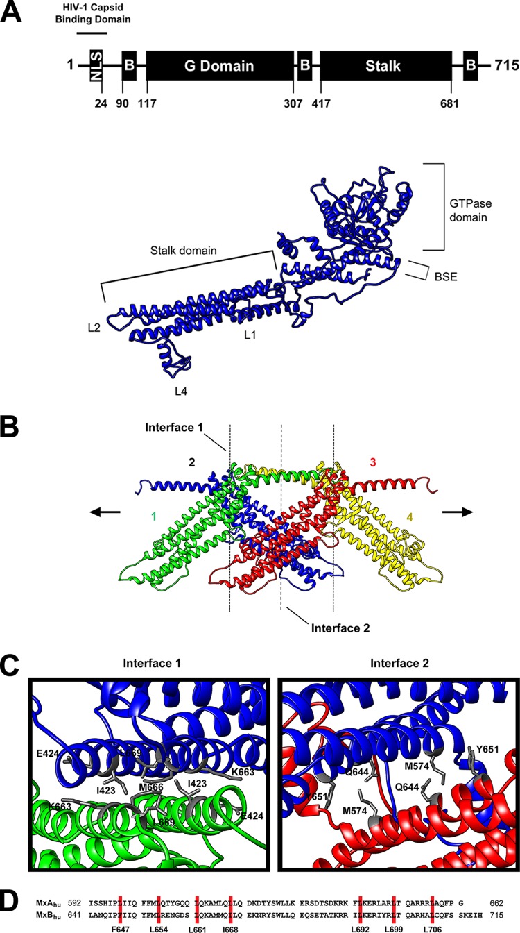
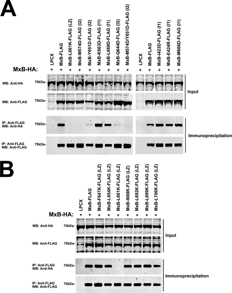
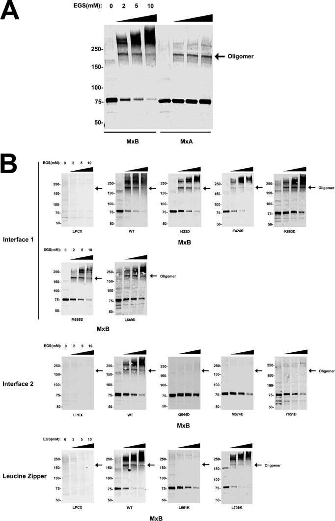
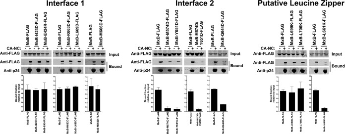
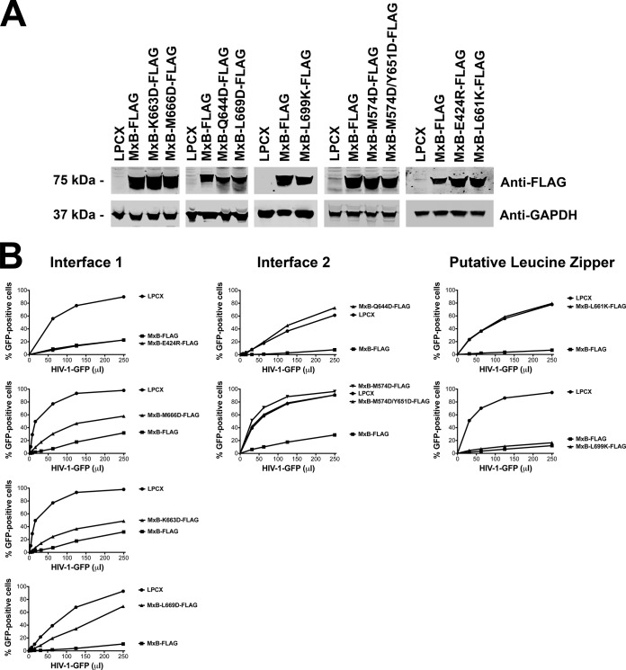
The alpha interferon (IFN-α)-inducible restriction factor myxovirus B (MxB) blocks HIV-1 infection after reverse transcription but prior to integration. MxB binds to the HIV-1 core, which is composed of capsid protein, and this interaction leads to inhibition of the uncoating process of HIV-1. Previous studies suggested that HIV-1 restriction by MxB requires binding to capsid. This work tests the hypothesis that MxB oligomerization is important for the ability of MxB to bind to the HIV-1 core. For this purpose, we modeled the structure of MxB using the published tertiary structure of MxA. The modeled structure of MxB guided our mutagenic studies and led to the discovery of several MxB variants that lose the capacity to oligomerize. In agreement with our hypothesis, MxB variants that lost the oligomerization capacity also lost the ability to bind to the HIV-1 core. MxB variants deficient for oligomerization were not able to block HIV-1 infection. Overall, our work showed that oligomerization is required for the ability of MxB to bind to the HIV-1 core and block HIV-1 infection.

Importance: MxB is a novel restriction factor that blocks infection of HIV-1. MxB is inducible by IFN-α, particularly in T cells. The current work studies the oligomerization determinants of MxB and carefully explores the contribution of oligomerization to capsid binding and restriction. This work takes advantage of the current structure of MxA and models the structure of MxB, which is used to guide structure-function studies. This work leads to the conclusion that MxB oligomerization is important for HIV-1 capsid binding and restriction.


Figures

FIG 1
FIG 1
Predicted structure and oligomerization of MxB. (A) Predicted structural model of human MxB. The wild-type human MxB protein is depicted. The numbers of the amino acid residues at the boundaries of the MxB domains are indicated. The structure of MxB was modeled using the published tertiary structure of MxA (PDB accession number 3SZR) using I-Tasser and Chimera (version 1.9) software. The nuclear localization signal (NLS), stalk domain, GTPase domain (G Domain), and bundle-signaling element (B) are indicated. (B) Predicted oligomerization interactions between MxB monomers. The MxB monomer was modeled into the dynamin 1 higher-order structure formation. (C) Top-down view of interface 1 and interface 2 of the predicted MxB structure. (D) Sequence alignment of human MxA (MxAhu) and human MxB (MxBhu) showing the putative leucine zipper domain in red.
FIG 2
FIG 2
Contribution of interface 1, interface 2, and putative leucine zipper residues to MxB oligomerization. (A) The ability of MxB variants with mutations in interface 1 and interface 2 to oligomerize was tested in human 293T cells. For this purpose, 293T cells were transfected with plasmids expressing wild-type MxB-HA and MxB-FLAG mutants. Cells were lysed at 24 h posttransfection and analyzed by Western blotting using anti-HA and anti-FLAG antibodies (Input). Lysates were immunoprecipitated using anti-FLAG agarose beads, and immunoprecipitates were eluted using 3× FLAG peptide. Elutions were analyzed by Western blotting using anti-HA and anti-FLAG antibodies (Immunoprecipitation). Representative results from three independent experiments are shown. (B) The ability of MxB putative leucine zipper variants to oligomerize was tested as described in the legend to panel A. Experiments were repeated at least three times, and representative results are shown. WB, Western blotting; IP, immunoprecipitation; LZ, putative leucine zipper; I1, interface 1; I2, interface 2.
FIG 3
FIG 3
Oligomerization of MxB variants by chemical cross-linking. (A) Human 293T cells were transfected with plasmids expressing FLAG-tagged human MxB or MxA. Cells were lysed at 24 h posttransfection. Lysates were incubated with EGS at the indicated concentrations and analyzed by Western blotting using anti-FLAG antibodies. (B) Similarly, the oligomerization ability of FLAG-tagged MxB variants with mutations in interface 1, interface 2, and the leucine zipper was measured by cross-linking experiments. Arrows, the oligomeric form of the indicated protein; WT, wild type. Numbers on the left are molecular masses (in kilodaltons).
FIG 4
FIG 4
Contribution of interface 1, interface 2, and putative leucine zipper residues to the ability of MxB to bind to in vitro-assembled HIV-1 CA-NC complexes. The ability of MxB variants to bind in vitro-assembled HIV-1 CA-NC complexes was measured. 293T cells were transfected with plasmids expressing FLAG-tagged wild-type or mutant MxB proteins. At 24 h posttransfection, the cells were lysed. Subsequently, the lysates were incubated at room temperature for 1 h with in vitro-assembled HIV-1 CA-NC complexes. The mixtures were applied to a 70% (wt/vol) sucrose cushion and centrifuged. Input, the lysates analyzed by Western blotting before being applied to the 70% cushion. The input mixtures were analyzed by Western blotting using anti-FLAG antibodies. The pellet from the 70% sucrose cushion (Bound) was analyzed by Western blotting using anti-FLAG or anti-p24 antibodies. Similar results were obtained in three independent experiments, and the standard deviation for the bound fraction relative to the results for the input is shown.
FIG 5
FIG 5
Residues on interface 2 are important for HIV-1 restriction by MxB. (A) Wild-type and mutant MxB proteins were stably expressed in Cf2Th cells. Levels of expression in whole-cell extracts were measured by Western blotting using anti-FLAG antibodies. The protein load was assayed by Western blotting using anti-GAPDH (anti- glyceraldehyde-3-phosphate dehydrogenase) antibodies. (B) Cf2Th cells stably expressing wild-type and mutant MxB proteins were challenged with increasing amounts of HIV-1–GFP. At 48 h postinfection, the percentage of GFP-positive cells was measured by flow cytometry. As a control, Cf2Th cells stably transduced with the empty vector LPCX were challenged with HIV-1. Similar results were obtained in three independent experiments, and representative results are shown.

References

    1. Mitchell PS, Emerman M, Malik HS. 2013. An evolutionary perspective on the broad antiviral specificity of MxA. Curr Opin Microbiol 16:493–499. doi:10.1016/j.mib.2013.04.005. - DOI - PMC - PubMed
    1. Haller O, Kochs G. 2011. Human MxA protein: an interferon-induced dynamin-like GTPase with broad antiviral activity. J Interferon Cytokine Res 31:79–87. doi:10.1089/jir.2010.0076. - DOI - PubMed
    1. Melén K, Keskinen P, Ronni T, Sareneva T, Lounatmaa K, Julkunen I. 1996. Human MxB protein, an interferon-alpha-inducible GTPase, contains a nuclear targeting signal and is localized in the heterochromatin region beneath the nuclear envelope. J Biol Chem 271:23478–23486. doi:10.1074/jbc.271.38.23478. - DOI - PubMed
    1. Aebi M, Fäh J, Hurt N, Samuel CE, Thomis D, Bazzigher L, Pavlovic J, Haller O, Staeheli P. 1989. cDNA structures and regulation of two interferon-induced human Mx proteins. Mol Cell Biol 9:5062–5072. - PMC - PubMed
    1. Haller O. 2013. Dynamins are forever: MxB inhibits HIV-1. Cell Host Microbe 14:371–373. doi:10.1016/j.chom.2013.10.002. - DOI - PubMed

Publication types

MeSH terms

Substances

Associated data