Review
. 2015 Sep 4;10(9):1636-50.
doi: 10.2215/CJN.06230614. Epub 2015 Jan 7.

Molecules Great and Small: The Complement System

Affiliations
Review

Molecules Great and Small: The Complement System

Douglas R Mathern et al. Clin J Am Soc Nephrol. .

Abstract

The complement cascade, traditionally considered an effector arm of innate immunity required for host defense against pathogens, is now recognized as a crucial pathogenic mediator of various kidney diseases. Complement components produced by the liver and circulating in the plasma undergo activation through the classical and/or mannose-binding lectin pathways to mediate anti-HLA antibody-initiated kidney transplant rejection and autoantibody-initiated GN, the latter including membranous glomerulopathy, antiglomerular basement membrane disease, and lupus nephritis. Inherited and/or acquired abnormalities of complement regulators, which requisitely limit restraint on alternative pathway complement activation, contribute to the pathogenesis of the C3 nephropathies and atypical hemolytic uremic syndrome. Increasing evidence links complement produced by endothelial cells and/or tubular cells to the pathogenesis of kidney ischemia-reperfusion injury and progressive kidney fibrosis. Data emerging since the mid-2000s additionally show that immune cells, including T cells and antigen-presenting cells, produce alternative pathway complement components during cognate interactions. The subsequent local complement activation yields production of the anaphylatoxins C3a and C5a, which bind to their respective receptors (C3aR and C5aR) on both partners to augment effector T-cell proliferation and survival, while simultaneously inhibiting regulatory T-cell induction and function. This immune cell-derived complement enhances pathogenic alloreactive T-cell immunity that results in transplant rejection and likely contributes to the pathogenesis of other T cell-mediated kidney diseases. C5a/C5aR ligations on neutrophils have additionally been shown to contribute to vascular inflammation in models of ANCA-mediated renal vasculitis. New translational immunology efforts along with the development of pharmacologic agents that block human complement components and receptors now permit testing of the intriguing concept that targeting complement in patients with an assortment of kidney diseases has the potential to abrogate disease progression and improve patient health.

Keywords: GN; complement; immunology; transplantation.


Figures

Figure 1.
Figure 1.
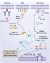
Overview of the complement cascade. The complement cascade can be initiated by three pathways: (1) the classical pathway, (2) the mannose-binding lectin (MBL) pathway, and (3) the alternative pathway. The resultant C3 convertases can continuously cleave C3; however, after they are generated, the alternative pathway C3 convertase dominates in amplifying production of C3b (green looping arrow). The C3 convertases associate with an additional C3b to form the C5 convertases, which cleave C5 to C5a+C5b. C5b recruits C6, C7, C8, and 10–16 C9 molecules to generate the terminal membrane attack complex (MAC), which inserts pores into cell membranes to induce cell lysis. C3a and C5a are potent signaling molecules, which through their G protein–coupled receptors C3aR and C5aR, respectively, can promote inflammation, chemoattraction of leukocytes, vasodilation, cytokine and chemokine release, and activation of adaptive immunity. fB, factor B; fD, factor D; fP, factor P; MASP, mannose-binding lectin-associated serine protease.
Figure 2.
Figure 2.
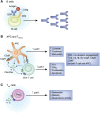
Complement and adaptive immunity. (A) B cells express complement receptor 2 (CR2; CD21), which binds to C3dg, a C3 breakdown product that functions as an opsonin. C3dg-coated antigens recognized by antigen-specific B-cell receptors plus CR2 initiate engulfment and lower the threshold of B-cell activation, promoting antibody production. (B) Cognate interactions between T cells and antigen-presenting cells (APCs) (T-cell receptor [TCR] +CD28/80/86 or CD40/154) upregulate the expression of multiple alternative pathway components, including C3, C5, fB, fD, C3aR, and C5aR, while simultaneously downregulating decay accelerating factor (DAF) expression (lifting restraint on complement activation). Locally produced C3a and C5a act in an autocrine/paracrine manner through AKT signaling to promote maturation and cytokine production of APCs, Th1/IFN-γ expression, increased proliferation, and decreased apoptosis of T cells. (C) Regulatory T-cell generation, stability, and suppressive function are decreased by C3a and C5a signaling-induced AKT signaling, which impairs nuclear translocation of Foxo1, a transcription factor for FoxP3. AKT, phosphokinase B; pAKT, phosphorylated phosphokinase B; BCR, B cell receptor; iTreg, murine-induced regulatory T cell.
Figure 3.
Figure 3.
Complement regulation. (A–D) Schematics depicting decay accelerating activity of (A and C) decay accelerating factor (DAF)/CD55 and factor H (fH), (B) CD59, which inhibits membrane attack complex formation, and (D) Membrane Cofactor Protein (MCP)/CD46 and fH, which display cofactor activity for factor I (fI). Cofactor-mediated fI activity irreversibly cleaves C3b to iC3b and subsequently cleaves iC3b to C3c and C3dg. (E) Schematic depicting the mechanism of C1-inhibitor (C1-inh), a protease that inactivates C1r, C1s, and mannose-binding lectin-associated serine proteases (MASPs), irreversibly preventing reformation of the classical and mannose-binding lectin (MBL) pathways initiating complexes. C1-inh also inhibits the kallikrein-kinin and coagulation cascades, two other mechanisms of complement activation.
Figure 4.
Figure 4.
Mechanisms through which complement mediates antibody-initiated kidney diseases. (A) Complement and membranous nephropathy (MN): autoantibodies reactive to podocyte-expressed phospholipase A2 receptor 1 (PLA2R1) activate complement through the mannose-binding lectin (MBL) pathway. Cascade amplification promotes membrane attack complex (MAC) formation, which induces sublytic signaling and dedifferentiation of the podocyte to impair its filtration capacity, leading to proteinuria. (B) Complement and immune complex disease: circulating or in situ–formed immune complexes from kidney disease initiated by lupus, streptococcal infections, and cryoglobulinemia are deposited in the subepithelial and/or subendothelial space of the glomerulus. Classical pathway activation induces inflammation and recruitment of neutrophils and macrophages (MΦ), promoting tissue injury. (C) Complement and ANCA vasculitis. Cytokine-induced neutrophil activation results in surface expression of myeloperoxidase (MPO; among other cytoplasmic antigens). On binding to ANCA and cross-linking FcR, local complement activation results in C5a production, which induces vascular inflammation by ligating its receptor C5aR. GBM, glomerular basement membrane; PMN, polymorphonuclear leukocyte.
Figure 5.
Figure 5.
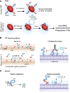
Kidney diseases caused by abnormalities in complement regulation. (A, upper panel) Healthy red blood cells (RBCs) express CD59 and decay accelerating factor (DAF), preventing complement activation on their surfaces. (A, lower panel) Paroxysmal nocturnal hemoglobinuria (PNH) is caused by mutation of the X-linked phosphatidylinositol glycan anchor biosynthesis, class A gene, resulting in a loss of glycophosphatidylinositol (GPI) anchor biosynthesis that prevents surface expression of GPI-anchored DAF (CD55) and CD59. An inability to regulate alternative pathway activation/amplification at the C3 convertase step (DAF; amplification loop) and prevent MAC formation (CD59), results in spontaneous RBC lysis (membrane attack complex [MAC]). (B) C3 nephropathies. Factor H (fH) normally binds to polyanionic glycosaminoglycans on glomerular basement membranes (GBMs) to restrain complement activation (Normal). Mutations in the C-terminal region of fH (depicted as red regions of the protein) impair fH’s physiologic ability to bind to the basement membrane, lifting restraint on local complement activation and contributing to glomerular inflammation and injury (mutant fH). C3 nephritic factors (right panel) can promote stability of C3, and properdin (factor P [fP]) can stabilize the C3bBb C3 convertase or inhibit fH-mediated decay/degradation, lifting restraint on C3 convertase-mediated amplification (looping arrow). (C) Atypical hemolytic uremic syndrome (aHUS). Among several mechanisms, mutations in fH that impair its ability to bind to basement membranes (red regions of protein) permit complement regulation in the fluid phase (plasma regulation; left panel) but prevent local complement regulation on surfaces (right panel), predisposing to vascular inflammation and hemolysis.
Figure 6.
Figure 6.
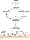
Complement in ischemia-reperfusion (IR) injury. IR injury results from tissue hypoxia and reperfusion, promoting reactive oxygen species (ROS) production, which causes endothelial injury. Inflammation drives Toll-like receptor (TLR)–mediated signaling and cytokine production as well as complement production and activation, which drives additional cytokine and chemokine production and immune cell recruitment through C3aR and C5aR signaling. DAMP, damage associated molecular pattern; HMGB1, high mobility group protein B1.
Figure 7.
Figure 7.
Chronic kidney injury and fibrosis. Chronic injury caused by toxins or transplant-related immune responses upregulates complement production and activation within the kidney. C3 has been implicated in the activation of the renin-angiotensin system and the promotion of the epithelial-to-mesenchymal transition, including fibrosis. IR, ischemia reperfusion; RAS, renin-angiotensin system.

References

    1. Cochrane CG, Unanue ER, Dixon FJ: A role of polymorphonuclear leukocytes and complement in nephrotoxic nephritis. J Exp Med 122: 99–116, 1965 - PMC - PubMed
    1. Brooimans RA, Stegmann AP, van Dorp WT, van der Ark AA, van der Woude FJ, van Es LA, Daha MR: Interleukin 2 mediates stimulation of complement C3 biosynthesis in human proximal tubular epithelial cells. J Clin Invest 88: 379–384, 1991 - PMC - PubMed
    1. Goodrum KJ: Complement component C3 secretion by mouse macrophage-like cell lines. J Leukoc Biol 41: 295–301, 1987 - PubMed
    1. Ricklin D, Hajishengallis G, Yang K, Lambris JD: Complement: A key system for immune surveillance and homeostasis. Nat Immunol 11: 785–797, 2010 - PMC - PubMed
    1. Diebolder CA, Beurskens FJ, de Jong RN, Koning RI, Strumane K, Lindorfer MA, Voorhorst M, Ugurlar D, Rosati S, Heck AJ, van de Winkel JG, Wilson IA, Koster AJ, Taylor RP, Saphire EO, Burton DR, Schuurman J, Gros P, Parren PW: Complement is activated by IgG hexamers assembled at the cell surface. Science 343: 1260–1263, 2014 - PMC - PubMed

Publication types

MeSH terms

Substances