. 2015 Feb 20;290(8):4801-4812.
doi: 10.1074/jbc.M114.605881. Epub 2015 Jan 7.

Phosphorylation of TET proteins is regulated via O-GlcNAcylation by the O-linked N-acetylglucosamine transferase (OGT)

Affiliations

Phosphorylation of TET proteins is regulated via O-GlcNAcylation by the O-linked N-acetylglucosamine transferase (OGT)

Christina Bauer et al. J Biol Chem. .

Abstract

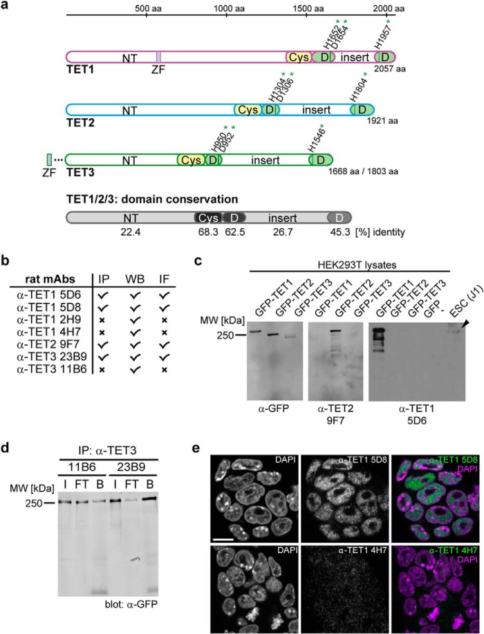
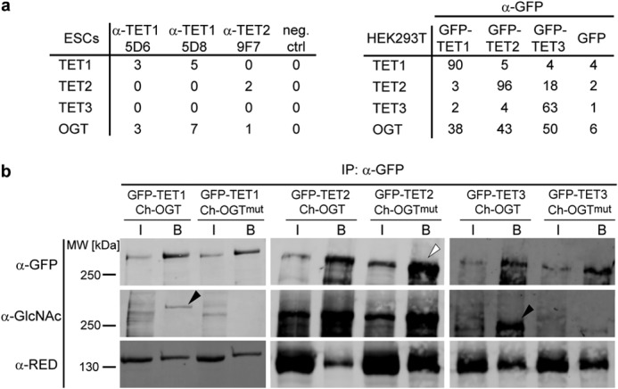
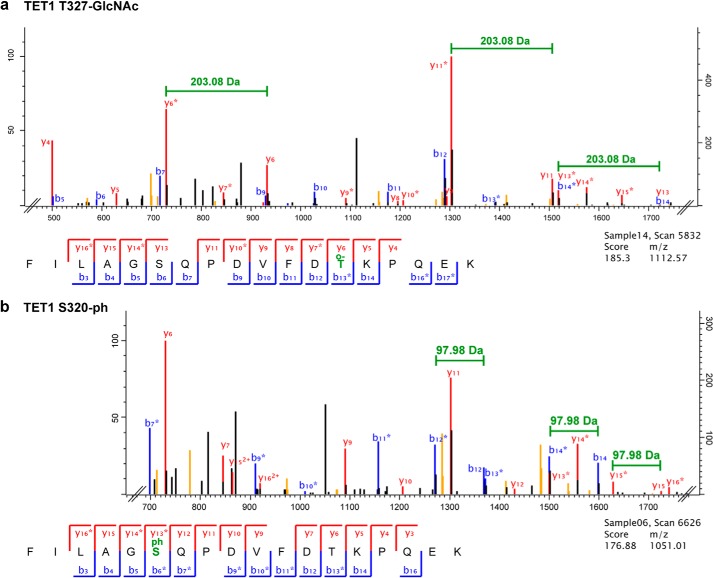
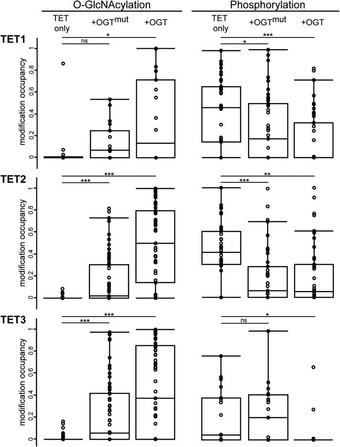
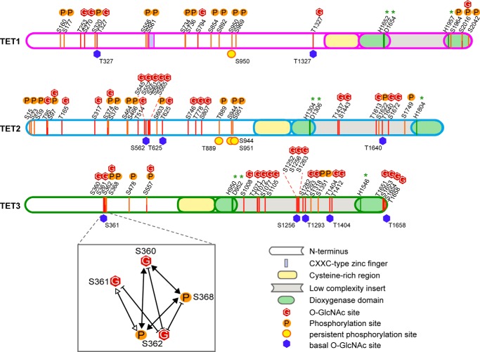
TET proteins oxidize 5-methylcytosine to 5-hydroxymethylcytosine, 5-formylcytosine, and 5-carboxylcytosine and thus provide a possible means for active DNA demethylation in mammals. Although their catalytic mechanism is well characterized and the catalytic dioxygenase domain is highly conserved, the function of the regulatory regions (the N terminus and the low-complexity insert between the two parts of the dioxygenase domains) is only poorly understood. Here, we demonstrate that TET proteins are subject to a variety of post-translational modifications that mostly occur at these regulatory regions. We mapped TET modification sites at amino acid resolution and show for the first time that TET1, TET2, and TET3 are highly phosphorylated. The O-linked GlcNAc transferase, which we identified as a strong interactor with all three TET proteins, catalyzes the addition of a GlcNAc group to serine and threonine residues of TET proteins and thereby decreases both the number of phosphorylation sites and site occupancy. Interestingly, the different TET proteins display unique post-translational modification patterns, and some modifications occur in distinct combinations. In summary, our results provide a novel potential mechanism for TET protein regulation based on a dynamic interplay of phosphorylation and O-GlcNAcylation at the N terminus and the low-complexity insert region. Our data suggest strong cross-talk between the modification sites that could allow rapid adaption of TET protein localization, activity, or targeting due to changing environmental conditions as well as in response to external stimuli.

Keywords: 5-Hydroxymethylcytosine (5-hmC); Dioxygenase; Epigenetics; O-Linked N-Acetylglucosamine (O-GlcNAc); OGT; Phosphorylation; Post-translational Modification (PTM); TET Proteins.


Figures

FIGURE 1.
FIGURE 1.
Generation of anti-TET monoclonal antibodies. a, schematic representation of the domain architecture of the three murine TET proteins. The catalytic dioxygenase domain (D) is split in two parts, separated by a presumably unstructured low-complexity insert (8), and is N-terminally preceded by a cysteine-rich region (Cys). The Fe(II)-binding residues are marked with green asterisks. The N terminus (NT) of TET1 contains a CXXC-type zinc finger (ZF). TET3 exists in two isoforms, one with a zinc finger and one without (41). The mean percent identity of the single domains of TET1, TET2, and TET3 is represented by different shades of gray and was calculated with Clustal 2.1 (59). aa, amino acids. b, overview of the generated anti-TET monoclonal antibodies (mAbs) and their possible applications. IP, immunoprecipitation; WB, Western blotting; IF, immunofluorescence; x, antibody not suited for the indicated application. c, example of Western blot analysis of two anti-TET antibodies with an anti-GFP antibody as a positive control. The antibodies detected only their target protein, but not the other two TET proteins. The WT protein from mESC whole cell lysates was also detected specifically (black arrowhead). d, example of an immunoprecipitation experiment with the indicated anti-TET3 antibodies. Clone 23B9 efficiently precipitated TET3 compared with clone 11B6. Western blot analysis was performed with an anti-GFP antibody. I, input; FT, flow-through; B, bound). e, immunofluorescence staining of mESCs with anti-TET1 antibodies (clones 5D8 and 4H7) and DAPI as a DNA counterstain. Whereas clone 5D8 showed a clear nuclear pattern, clone 4H7 displayed only a weak and diffuse signal. Confocal imaging was performed with a Leica TCS SP5 confocal laser scanning microscope with a ×63/1.4 numerical aperture Plan-Apochromat oil immersion objective. Scale bar = 5 μm.
FIGURE 2.
FIGURE 2.
TET proteins interact with OGT and are O-GlcNAcylated. a, number of unique peptides detected in immunoprecipitation experiments followed by LC-MS/MS. Left, immunoprecipitation of endogenous TET1 or TET2 with the indicated antibodies. Protein G beads without antibody were used as a negative control (neg. ctrl). Right, immunoprecipitation of GFP-tagged TET1, TET2, or TET3 expressed in HEK293T cells. Pulldown of GFP served as a negative control. b, Western blot analysis of TET1, TET2, and TET3 specifically enriched with GFP-Trap. Upon coexpression of active OGT, the O-GlcNAcylation signal increased for TET1 and TET3 (black arrowheads) compared with coexpression of catalytically inactive OGTmut. For TET2, protein levels in the OGTmut samples were higher (white arrowhead), whereas the O-GlcNAc signal remained constant, suggesting a higher proportion of O-GlcNAcylated TET2 in the OGT sample. Interaction between TET proteins and OGT was independent of OGT activity. Anti-RED antibody (60) detected the coexpressed Cherry (Ch)-tagged OGT. IP, immunoprecipitation; I, input; B, bound.
FIGURE 3.
FIGURE 3.
Exemplary MS/MS spectra of modified TET1 peptides. a, MS/MS spectrum of a TET1 peptide modified with O-GlcNAc (o-) at the threonine residue. O-GlcNAcylation is characterized by a neutral loss of 203.8 Da as indicated. y ions are depicted in red, and b ions are depicted in blue. Labeling of neutral losses of H2O or NH3 (orange peaks) has been removed for clarity. Fully annotated spectra are provided in supplemental Data S2 and S3. b, MS/MS spectrum of the same TET1 peptide phosphorylated (-ph) at the serine residue. Phosphorylated ions show a neutral loss of 97.98 Da as indicated. y ions are depicted in red, b ions are depicted in blue. Labeling of neutral losses of H2O or NH3 (orange peaks) has been removed for clarity. Fully annotated spectra are provided in supplemental Data S2 and S3.
FIGURE 4.
FIGURE 4.
TET phosphorylation is reduced upon O-GlcNAcylation. Box plots depict the distribution of O-GlcNAc and phosphorylation occupancy in the three conditions: expression of TET protein only, coexpression of OGTmut, and coexpression of OGT. Missing values have been substituted with an occupancy of 0.005, with 0.001 being the lowest measured occupancy. Mean occupancies of single sites are provided in Tables 1–3. *, p < 0.05 (Student's t test); **, p < 0.01; ***, p < 0.001; ns, not significant.
FIGURE 5.
FIGURE 5.
N termini and insert regions of TET proteins are densely modified. Shown are a schematic and scaled mapping of all TET phosphorylation and O-GlcNAcylation sites in the protein sequence. Modifications are found mostly in the N terminus and insert region and rarely occur at the same residue. Residue numbering refers to the murine protein sequences specified in supplemental Data S1. Green asterisks indicate catalytic Fe(II)-binding residues. Basal O-GlcNAc sites occur without any coexpression of OGT or OGTmut; persistent phosphorylation sites show high occupancy despite an increase in O-GlcNAcylation. An example of the PTM cross-talk on TET proteins is shown for TET3 Ser-360/Ser-361/Ser-362/Ser-368. White arrowheads, two co-occurring modifications; black arrowheads, three co-occurring modifications; blunt arrows, mutual exclusivity.

References

    1. Goll M. G., Bestor T. H. (2005) Eukaryotic cytosine methyltransferases. Annu. Rev. Biochem. 74, 481–514 - PubMed
    1. Suzuki M. M., Bird A. (2008) DNA methylation landscapes: provocative insights from epigenomics. Nat. Rev. Genet. 9, 465–476 - PubMed
    1. Tahiliani M., Koh K. P., Shen Y., Pastor W. A., Bandukwala H., Brudno Y., Agarwal S., Iyer L. M., Liu D. R., Aravind L., Rao A. (2009) Conversion of 5-methylcytosine to 5-hydroxymethylcytosine in mammalian DNA by MLL partner TET1. Science 324, 930–935 - PMC - PubMed
    1. Ito S., Shen L., Dai Q., Wu S. C., Collins L. B., Swenberg J. A., He C., Zhang Y. (2011) Tet proteins can convert 5-methylcytosine to 5-formylcytosine and 5-carboxylcytosine. Science 333, 1300–1303 - PMC - PubMed
    1. He Y. F., Li B. Z., Li Z., Liu P., Wang Y., Tang Q., Ding J., Jia Y., Chen Z., Li L., Sun Y., Li X., Dai Q., Song C. X., Zhang K., He C., Xu G. L. (2011) Tet-mediated formation of 5-carboxylcytosine and its excision by TDG in mammalian DNA. Science 333, 1303–1307 - PMC - PubMed

Publication types

MeSH terms