. 2015 Feb 27;290(9):5226-39.
doi: 10.1074/jbc.M114.626382. Epub 2015 Jan 7.

Crystal structures capture three states in the catalytic cycle of a pyridoxal phosphate (PLP) synthase

Affiliations

Crystal structures capture three states in the catalytic cycle of a pyridoxal phosphate (PLP) synthase

Amber Marie Smith et al. J Biol Chem. .

Abstract

PLP synthase (PLPS) is a remarkable single-enzyme biosynthetic pathway that produces pyridoxal 5'-phosphate (PLP) from glutamine, ribose 5-phosphate, and glyceraldehyde 3-phosphate. The intact enzyme includes 12 synthase and 12 glutaminase subunits. PLP synthesis occurs in the synthase active site by a complicated mechanism involving at least two covalent intermediates at a catalytic lysine. The first intermediate forms with ribose 5-phosphate. The glutaminase subunit is a glutamine amidotransferase that hydrolyzes glutamine and channels ammonia to the synthase active site. Ammonia attack on the first covalent intermediate forms the second intermediate. Glyceraldehyde 3-phosphate reacts with the second intermediate to form PLP. To investigate the mechanism of the synthase subunit, crystal structures were obtained for three intermediate states of the Geobacillus stearothermophilus intact PLPS or its synthase subunit. The structures capture the synthase active site at three distinct steps in its complicated catalytic cycle, provide insights into the elusive mechanism, and illustrate the coordinated motions within the synthase subunit that separate the catalytic states. In the intact PLPS with a Michaelis-like intermediate in the glutaminase active site, the first covalent intermediate of the synthase is fully sequestered within the enzyme by the ordering of a generally disordered 20-residue C-terminal tail. Following addition of ammonia, the synthase active site opens and admits the Lys-149 side chain, which participates in formation of the second intermediate and PLP. Roles are identified for conserved Asp-24 in the formation of the first intermediate and for conserved Arg-147 in the conversion of the first to the second intermediate.

Keywords: Enzyme Structure; Glutaminase; Pyridoxal Phosphate; Vitamin; X-ray Crystallography.


Figures

FIGURE 1.
FIGURE 1.
PLP biosynthesis by the R5P-dependent pathway (PdxS/PdxT) in G. stearothermophilus. A, glutaminase reaction of PdxT. B, synthase reactions of PdxS. I1 forms upon R5P addition to Lys-81, I2 upon ammonia addition to I1, and PLP upon G3P addition to I2. A proposed structure (16) for the I2 chromophore is shown.
FIGURE 2.
FIGURE 2.
Alternative nitrogen sources for PLPS. I2 chromophore formation is compared for wild type PLPS, PLPS with an inactivated glutaminase (PLPS/H169NT), and the synthase subunit (PdxS). The nitrogen sources are glutamine or NH3 from (NH4)2SO4. Exogenous NH3 can substitute for PdxT and glutamine. Data on a relative scale are averages of triplicate reactions.
FIGURE 3.
FIGURE 3.
PdxS·I1. R5P forms I1 at Lys-81 and extends across the (β/α)8 barrel. Under long incubation, Lys-149 forms an off-pathway adduct with R5P C2 outside the active site. Electron density is shown for Lys-81-I1 and Lys-149-R5P (FoFc omit contoured in green at 2.5 σ). Inset, salt bridge network (orange/yellow dashes) at the Lys-149–5RP phosphate superposed with the Saccharomyces cerevisiae synthase-PLP complex (Protein Data Bank code 3O05 (11)). Key amino acids are shown as sticks in atomic coloring (blue, N; red, O; orange, P) with cyan, C, for PdxS residues hydrogen-bonded (yellow dashes) to ligands (white C for I1 and R5P; green C for PLP). Residues in the extensive salt bridge network are also shown (orange C). The boundaries of disordered regions at residues 47–56 and the C terminus (271–294) are shown as cyan spheres.
FIGURE 4.
FIGURE 4.
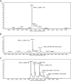
Formation of I1 and I2 in PdxS. A, deconvoluted electrospray injection (ESI) mass spectrum showing complete conversion of PdxS to PdxS·I1 upon a 15-min incubation with R5P. The calculated mass of the PdxS free enzyme is 31,849.6 Da. B, accumulation of a second adduct (+ 212 Da) upon a 48-h incubation with an excess of R5P. Calculated masses are shown on the spectra. C, deconvoluted electrospray injection mass spectrum showing accumulation of PdxS·I2 upon a 15-min incubation with R5P and (NH4)2SO4. A mixture of PdxS·I2, PdxS·I1, PdxS·I2/Lys-149-R5P, PdxS·I1/Lys-149-R5P, and free enzyme was detected.
FIGURE 5.
FIGURE 5.
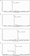
Amino acids critical to accumulation of PdxS·I1 and PdxS·I2. A, deconvoluted ESI mass spectrum for PdxS/D24N after a 15-min incubation with R5P. B, deconvoluted ESI mass spectrum for PdxS/D24A after a 15-min incubation with R5P. No mass corresponding to I1 (+ 212 Da) accumulates for either variant at amino acid 24. C, deconvoluted ESI mass spectrum for PdxS/R147Q-I1 after a 15-min incubation with R5P and (NH4)2SO4. D, deconvoluted ESI mass spectrum for PdxS/R147A after a 15-min incubation with R5P and (NH4)2SO4. Accumulation of I1 is slightly reduced in PdxS/R147A, as a small amount of the free enzyme remained. No mass corresponding to PdxS·I2 (+ Da) was detected for either variant at amino acid 147.
FIGURE 6.
FIGURE 6.
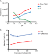
I2 accumulation. A, effect of pH on distribution of PdxS species after incubation with R5P and (NH4)2SO4. Peak areas are from deconvoluted ESI mass spectra. B, effect of (NH4)2SO4 concentration on distribution of PdxS species after incubation with R5P and (NH4)2SO4. I2 accumulation is compared with the total modified PdxS, based on peak areas from deconvoluted ESI mass spectra. No conditions were identified in which I2 was the predominant form of PdxS.
FIGURE 7.
FIGURE 7.
PdxS·I1/I2. Electron density for intermediates I1 (left) and I2 (right) at Lys-81 (FoFc omit density in green contoured at 2.5 σ). Intermediates (white C) and key amino acids (orange C) are in stick form.
FIGURE 8.
FIGURE 8.
PLPS·I1·Glu. A, 24 subunit intact PLPS (red, pink PdxS; light, dark gray PdxT). The PdxS-PdxT protomer is shown at right (yellow PdxT active site and PdxS C-terminal tail, green glutamyl thioester, and gray I1). B, PdxT active site shown in stereo. The H169N substitution trapped the glutamyl-thioester intermediate at Cys-78 (FoFc omit density contoured at 2.5 σ in green). C, closed synthase subunit. The C-terminal tail (yellow) and helices α2a and α8′ cover I1 and the KGEPG loop (purple). Electron density is shown for I1 at Lys-81 and in stereo for the C-terminal tail (FoFc omit density contoured at 2.5 σ in green; 2FoFc density contoured at 1 σ in blue). Inset, interactions of helices α2a and α8′ (green), the KGEPG loop (purple) with the C-terminal tail yellow) and with I1 (white).
FIGURE 9.
FIGURE 9.
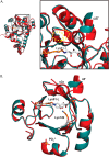
Conformational changes in the synthase active site. A, closure around I1 in PLPS·I1·Glu. Superposition of PLPS·I1·Glu (red) and PdxS·I1/I2 (teal) illustrates how synthase closure forms helix α2a and shifts helix α8′ toward I1 relative to its position in the open structures, here represented by PdxS·I1/I2. The disorder of amino acids 48–56 in PdxS·I1/I2 is indicated by yellow dashes. B, comparison of Lys-149 positions. Lys-149, located on the invariant KGEPG loop, points into the active site in PdxS·I1/I2 (teal) and outside the active site in all other structures, here illustrated by superposition with PLPS·I1·Glu (red). The KGEPG loop also shifts with movement of Lys-149 into the active site. A phosphate ion is shown in the secondary site.

Similar articles

Cited by

References

    1. Eliot A. C., Kirsch J. F. (2004) Pyridoxal phosphate enzymes: mechanistic, structural, and evolutionary considerations. Annu. Rev. Biochem. 73, 383–415 - PubMed
    1. Christen P., Mehta P. K. (2001) From cofactor to enzymes. The molecular evolution of pyridoxal-5′-phosphate-dependent enzymes. Chem. Rec. 1, 436–447 - PubMed
    1. Jain S. K., Lim G. (2001) Pyridoxine and pyridoxamine inhibits superoxide radicals and prevents lipid peroxidation, protein glycosylation, and (Na+ + K+)-ATPase activity reduction in high glucose-treated human erythrocytes. Free Radic. Biol. Med. 30, 232–237 - PubMed
    1. Bilski P., Li M. Y., Ehrenshaft M., Daub M. E., Chignell C. F. (2000) Vitamin B6 (pyridoxine) and its derivatives are efficient singlet oxygen quenchers and potential fungal antioxidants. Photochem. Photobiol. 71, 129–134 - PubMed
    1. Ehrenshaft M., Jenns A. E., Chung K. R., Daub M. E. (1998) SOR1, a gene required for photosensitizer and singlet oxygen resistance in Cercospora fungi, is highly conserved in divergent organisms. Mol. Cell 1, 603–609 - PubMed

Publication types

MeSH terms