Southwick osteotomy stabilised with external fixator
- PMID: 25568571
- PMCID: PMC4269534
- DOI: 10.5455/medarh.2014.68.353-355
Southwick osteotomy stabilised with external fixator
Abstract
Introduction: Epiphysiolysis of the femoral head is the most common accident occurring towards the end of pre-puberty and puberty growth.
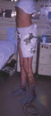
Case report: The author describes the experience in the treatment of chronic epiphysiolysis in two patients treated by Southwick osteotomy. The site is accessed by way of a 15-cm long lateral skin incision and the trochanteric region is reached through the layers. The osteotomy angles prepared beforehand on a thin aluminium model are used to mark the Southwick osteotomy site on the anterior and lateral sides at the level of the lesser trochanter. Before performing the trochanteric osteotomy, two Mitković convergent pins type M20 are applied distally and proximally, above the planned osteotomy site. A tenotomy of the iliopsas muscle is performed, and then the previously marked bone triangle is redissected up to three quarters of the width of the femur. The distal part of the femur is rotated inwards, so that the patella is turned towards the ceiling. The osteotomised fragments of the femur are adapted, repositioned and fixated by installing an external fixator on the previously placed pins. Two more pins are placed, one proximally and one distally, with a view to adequately stabilising the femur. The patient was mobile from day two after the surgery. If, after the surgery, the lead surgeon realises that there is a requirement to make a correction of 5, 10 and 15 degrees of the valgus, varus, anteversion or retroversion deformity, the correction shall be performed without surgically opening the patient, using the fixator pins.
Conclusion: After performing a Southwick osteotomy it is easier to adapt, reposition and fixate the osteotomised fragments of the femur using a fixator type M20. Adequate stability allows regaining mobility quickly, which in turn is the best prevention of chondrolysis of the hip. It is possible to make post-operative valgus, varus, anteversion and retroversion corrections of 5, 10 and 15 degrees without performing a surgery. Once the osteotomy is healed, the fixator type M20 is removed without any additional surgery.
Keywords: Southwick osteotomy; external fixator.
Conflict of interest statement
Figures


References
-
- Petković L. Novom Sadu, Novi Sad: Medicinski fakultet Univerziteta u; 2007. Bolni kuk i ultrazvučna dijagnostika u razvojnom dobu.
-
- Slavković S. Beograd: Monografija; 1986. Epizioliza proksimalne epifize femura.
-
- Slavković S. Neoperativno lečenje akutnog smicanja glave femura. Acta Orth Iugoslavica. 1992;1-2:21–25.
-
- Soutwick WO. Slipid Capital Femoral Epiphysis. J Bone J Surg. 1984;66-A:1151–1152. - PubMed
-
- Grubor P, Grubor M. Results of Application of Eexternal Fixation with Different Types of Fixators. Srp Arh Celok Lec. 2012 May-Jun;140(5-6):332–338. - PubMed
Publication types
MeSH terms
LinkOut - more resources
Full Text Sources
Other Literature Sources
