. 2015 Jan 8;11(1):e1004923.
doi: 10.1371/journal.pgen.1004923. eCollection 2015 Jan.

ASAR15, A cis-acting locus that controls chromosome-wide replication timing and stability of human chromosome 15

Affiliations

ASAR15, A cis-acting locus that controls chromosome-wide replication timing and stability of human chromosome 15

Nathan Donley et al. PLoS Genet. .

Abstract

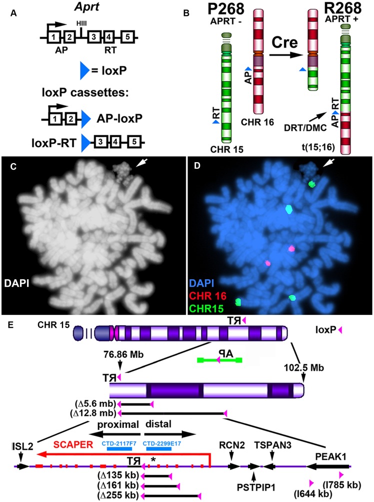
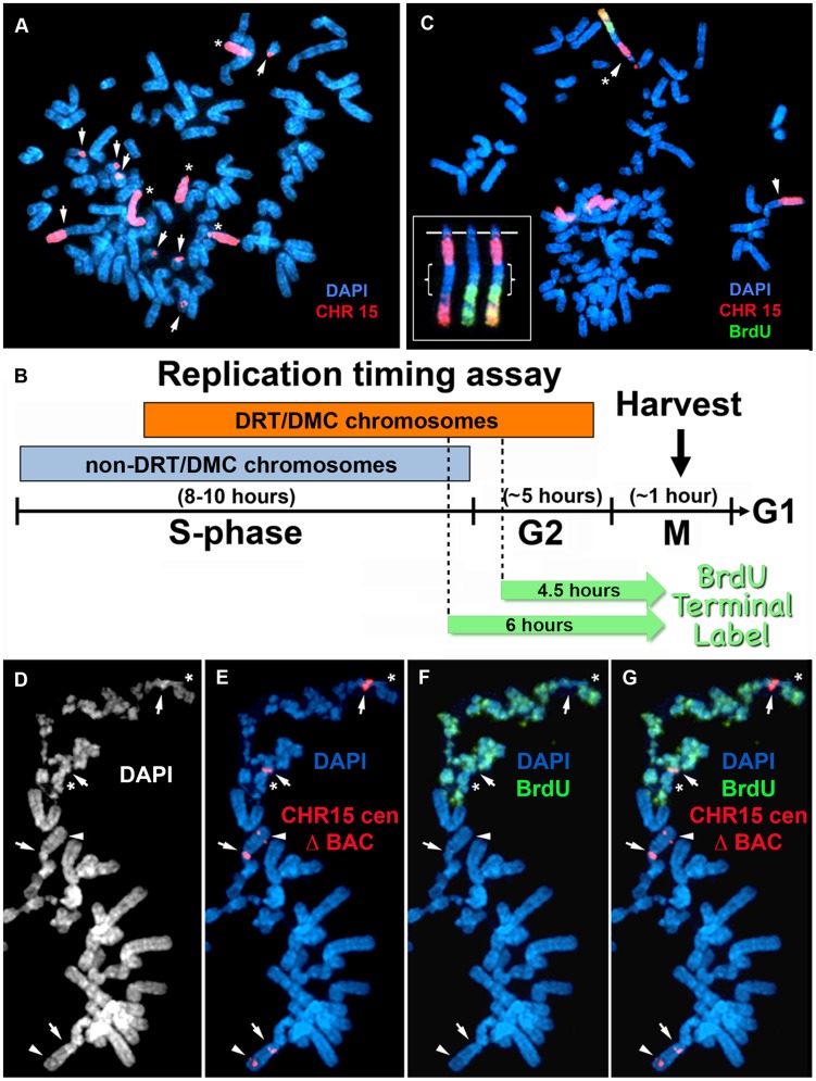
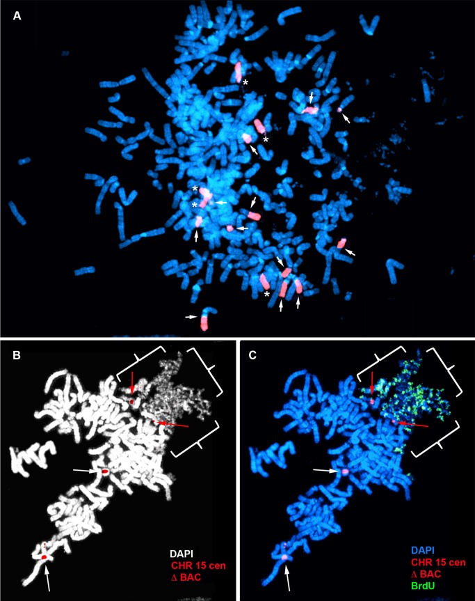
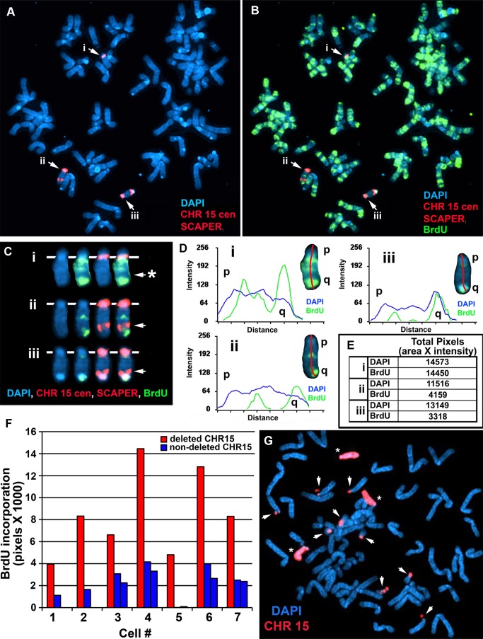
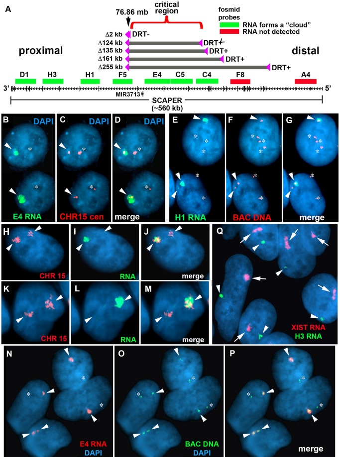
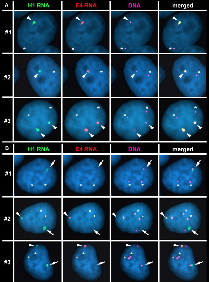
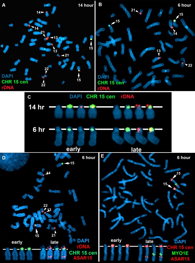
DNA replication initiates at multiple sites along each mammalian chromosome at different times during each S phase, following a temporal replication program. We have used a Cre/loxP-based strategy to identify cis-acting elements that control this replication-timing program on individual human chromosomes. In this report, we show that rearrangements at a complex locus at chromosome 15q24.3 result in delayed replication and structural instability of human chromosome 15. Characterization of this locus identified long, RNA transcripts that are retained in the nucleus and form a "cloud" on one homolog of chromosome 15. We also found that this locus displays asynchronous replication that is coordinated with other random monoallelic genes on chromosome 15. We have named this locus ASynchronous replication and Autosomal RNA on chromosome 15, or ASAR15. Previously, we found that disruption of the ASAR6 lincRNA gene results in delayed replication, delayed mitotic condensation and structural instability of human chromosome 6. Previous studies in the mouse found that deletion of the Xist gene, from the X chromosome in adult somatic cells, results in a delayed replication and instability phenotype that is indistinguishable from the phenotype caused by disruption of either ASAR6 or ASAR15. In addition, delayed replication and chromosome instability were detected following structural rearrangement of many different human or mouse chromosomes. These observations suggest that all mammalian chromosomes contain similar cis-acting loci. Thus, under this scenario, all mammalian chromosomes contain four distinct types of essential cis-acting elements: origins, telomeres, centromeres and "inactivation/stability centers", all functioning to promote proper replication, segregation and structural stability of each chromosome.


Conflict of interest statement

The authors have declared that no competing interests exist.

Figures

Figure 1
Figure 1. DRT/DMC on an engineered t (15;16).
A) Diagram of the Aprt-loxP cassettes, and the genomic organization of the mouse Aprt gene is shown. The 5′ portion of the Aprt gene was separated from the 3′ portion to generate the AP-loxP and loxP-RT cassettes. Each cassette contains a loxP site in the second intron of the Aprt gene, and each cassette was cloned separately into Lentiviral vectors. B) Illustration of the original loxP cassette integration sites in chromosomes 15 (green) and 16 (red) in P268 cells, and the balanced translocation, t (15;16), generated in R268 cells (see [17]). C and D) DMC on the chromosome 15 derivative of the t (15;16). R268 cells were harvested for mitotic cells, dropped on slides and processed for DNA FISH using chromosome 15 and 16 centromeric probes [CHR15 (green) and CHR16 (red)]. E) Diagram of chromosome 15 showing the orientation and integration site (∼76.86 mb) of the original loxP-3′RT cassette in P268 cells. P268 cells were infected with Lentiviral vectors containing either the AP-loxP or loxP-RT cassettes, and 18 pools of 5,000–10,000 infected clones isolated for each Lentivirus. The structure of the AP-loxP Lentivirus (in the opposite orientation; green), the extent of 5 distal deletions (Δ135 kb, Δ161 kb, Δ255 kb, Δ5.6 mb and Δ12.8 mb) and 2 inversions (I644 kb, and I785 kb), BACs (CTD-2117F7 and CTD-2299E17), and the protein-coding genes ISL2, SCAPER, RCN2, PSTPIP1, TSPAN3 and PEAK1 are indicated. The approximate location of the micro RNA gene MIR3713 is shown with an asterisk.
Figure 2
Figure 2. Chromosome rearrangements and delayed replication of a Cre/loxP-mediated deletion (∼135 kb) in chromosome 15.
A) Secondary rearrangements of chromosome 15. Δ268-4f cells were processed for DNA FISH using a chromosome 15 WCP, and the DNA was stained with DAPI. Rearrangements involving chromosome 15 are indicated with arrows. Non-rearranged chromosome 15 s are indicated with asterisks. B) Schematic diagram of the BrdU Terminal Label replication-timing assay . Cells were exposed to BrdU for either 4.5 or 6 hours, harvested for mitotic cells, and processed for BrdU incorporation and DNA FISH to identify chromosome 15. C) BrdU-WCP assay on cells containing an ∼135 kb distal deletion in chromosome 15. Δ268-4f cells were exposed to BrdU for 4.5 hours, harvested for mitotic cells, stained with an anti-BrdU antibody (green), and processed for DNA FISH with a chromosome 15 WCP (CHR 15; red). The DNA was stained with DAPI. Two different chromosome 15 secondary rearrangements are indicated with arrows. The inset shows the derivative chromosome 15 with the asterisk, with the BrdU staining and WCP shown in separate images. The brackets highlight the non-chromosome 15 DNA. D–G) BrdU-BAC assay on cells containing an ∼135 kb distal deletion in chromosome 15. Δ268-4c cells were exposed to BrdU for 4.5 hours, harvested for mitotic cells, stained with an anti-BrdU antibody (green), and processed for DNA FISH with a chromosome 15 centromeric probe (red) plus a BAC (CTD-2299E17; red) from the deleted region. The DNA was stained with DAPI (white in panel D or blue in panels E–G). The arrows mark the centromeric signals, and the arrowheads mark the BAC signals. The asterisks mark short arms of the deleted chromosome 15 s, which contain BrdU incorporation.
Figure 3
Figure 3. Chromosome rearrangements and delayed replication of a Cre/loxP-mediated deletion (∼5.6 mb) in chromosome 15.
A) Secondary rearrangements of chromosome 15. Mitotic Δ268-5d cells were processed for DNA FISH using a chromosome 15 WCP, and the chromosomal DNA was stained with DAPI. Rearrangements involving chromosome 15 are indicated with arrows. Non-rearranged chromosome 15 s are indicated with asterisks. B and C) BrdU-BAC replication assay on cells containing the ∼5.6 mb distal deletion in chromosome 15. Δ268-5c cells were exposed to BrdU for 4.5 hours, harvested for mitotic cells, stained with an antibody to BrdU (green), and processed for DNA FISH with a chromosome 15 centromeric probe (red) plus a BAC (CTD-2299E17; ΔBAC, red) from the deleted region. The DNA was stained with DAPI (white in panel B or blue in panel C). The red arrows mark the centromeric signals from two chromosomes with DRT/DMC. The white arrows mark the centromeric signals from two non-DRT/DMC chromosome 15 s. The brackets highlight the extreme “pulverization” of two deleted chromosome 15 s.
Figure 4
Figure 4. Delayed replication of chromosome 15 with an Cre/loxP-mediated ∼161 kb distal deletion.
A–F) Δ268-4 g cells were incubated with BrdU for 6 hours, harvested for mitotic cells, stained with an antibody to BrdU (green) and processed for DNA FISH using a chromosome 15 centromeric probe (red) plus BAC CTD-2299E17 (red). The chromosomal DNA was stained with DAPI (blue). A and B) A metaphase spread containing three chromosome 15 s (i, ii, and iii). C) The three chromosome 15 s from panel B were cut out and aligned showing the BrdU and FISH signals in separate images. The asterisk marks the location of the deletion in the chromosome marked i, and the arrows mark the location of the BAC hybridization signals on chromosomes ii and iii. D) Pixel intensity profiles of the BrdU incorporation (green), and DAPI (blue) staining along the three chromosome 15 s from panel B. E) The pixel intensity (average intensity x area) for each chromosome, i, ii, and iii, showing the total amount of BrdU incorporation or DAPI staining. F) Quantification of the BrdU incorporation in multiple cells. The red and blue bars represent deleted and non-deleted chromosome 15 s, respectively, in 7 different cells. G) Instability of chromosome 15 containing an ∼161 kb Cre-loxP deletion. Mitotic Δ268-4e cells were processed for DNA FISH with a chromosome 15 WCP, and the chromosomal DNA was stained with DAPI. Rearrangements involving chromosome 15 are indicated with arrows, and non-rearranged chromosome 15 s are indicated with asterisks.
Figure 5
Figure 5. Differential allelic expression of ASAR15.
A) A schematic diagram of SCAPER, MIR3713, RNA FISH probes, and five of the distal deletions in chromosome 15. The genomic location (in megabases), the exon-intron structure of SCAPER, the location of 9 fosmids [G248P87971D1 (D1), G248P81306H3 (H3), G248P82406H1 (H1), G248P87518F5 (F5), G248P82172E4 (E4), G248P8912C5 (C5), G248P89264C4 (C4), G248P88942F8 (F8), G248P80481A4 (A4)] used for RNA FISH, and the location of the five smallest distal deletions are shown. The fosmids that detect (green) or do not detect (red) RNA are indicated. B–D) Monoallelic expression of ASAR15 in HTD114 cells. HTD114 cells were subjected to RNA FISH using a fosmid (E4 RNA; green) to detect RNA. Images of the RNA hybridization signals were obtained, and the coordinates of individual cells were recorded. Slides were subsequently processed for DNA FISH using a chromosome 15 centromeric probe (CHR15 cen; red) and new images of the DNA hybridization were captured for the same cells. E–G) P268 cells were processed for RNA-DNA FISH using the H1 probe to detect RNA, plus BAC CTD-2299E17 to detect DNA. H-M) RNA-DNA FISH using a pool of fosmid probes (D1, H3, H1, F5, E4, C5, and C4) to detect RNA (green) and a chromosome 15 paint to detect DNA (red). Panels H–J and K-M represent two different cells with the images shown in separate or merged panels. The arrowheads mark the chromosome 15s. N–P) Primary HFFs were processed for RNA-DNA FISH using probe E4 to detect RNA, plus BAC CTD-2117F7 to detect DNA. B–J) The nuclear DNA was stained with DAPI. Arrowheads mark the location of RNA signals, and asterisks mark the location of the DNA signals that lack corresponding RNA signals. In regions of the slides where the FISH worked well, the D1, H3, H1, F5, E4, C5, and C4 probes detected a positive signal in>90% of the HTD114, P268 and HFF cells. Q) RNA FISH in female HDFs. Female HDF cells were processed for RNA FISH using the H1 ASAR15 probe (green) in combination with an XIST (red) probe. The arrows mark the large clouds of RNA detected by the XIST probe and the arrowheads mark hybridization signals detected by the ASAR15 probe. The DNA was stained with DAPI.
Figure 6
Figure 6. The Cre/loxP-mediated deletions occurred on the expressed allele of ASAR15.
P268 (panel A) and Δ268-4 g (panel B) cells were subjected to RNA FISH using the H1 (green) and E4 (red) probes to detect RNA. Images of the RNA hybridization signals were obtained, and the coordinates of individual cells were recorded; the slides were subsequently processed for DNA FISH (BACs CTD-2299E17 plus BAC-CTD-2117F7) and new images of the DNA hybridization were captured for the same cells. The DNA FISH step included an RNAase step, which eliminated the RNA FISH signals. The DNA FISH hybridization signal was pseudo-colored purple for clarity, and the nuclear DNA was stained with DAPI (blue). Representative images from three different P268 and Δ268-4 g cells (#1–3) are shown in each panel (A and B). The arrowheads mark the coincident sites of hybridization detected by both probes. The arrows mark the sites of hybridization with the H1 probe that was not coincident with a site of hybridization with the E4 probe in Δ268-4 g cells (panel B). The asterisks mark the sites of DNA hybridization that lacked corresponding RNA hybridization signals from either H1 or E4 probes.
Figure 7
Figure 7. Coordinated random asynchronous replication on chromosome 15.
A–C) ReTiSH assay on rDNA loci in PBLs. PBLs were labeled with BrdU for 14 (A) or 6 (B) hours, arrested in metaphase, and subjected to ReTiSH using an 18S rDNA probe (red). The chromosome 15 s were identified using a centromeric probe (green), and the chromosomal DNA was detected with DAPI. A and B) The DAPI images of the chromosomes were inverted and the banding patterns were used to identify all of the ReTiSH positive chromosomes. The arrows mark the chromosome 15 s, and the arrowheads mark the other four chromosomes containing rDNA clusters (13, 14, 21, and 22). C) The ReTiSH signals for the rDNA (red) and chromosome 15 centromeric (green) probes from the 14 (panel A) and 6 (panel B) hour time points are shown. The early and late replicating chromosome 15 s are indicated for the 6 hour time point. D) ReTiSH assay using an ASAR15 BAC (CTD-2299E17; red), an rDNA probe (red), and a chromosome 15 centromeric probe (green). The ASAR15 and rDNA probes show hybridization signals to the same chromosome 15 homolog at the 6 hour time point. E) ReTiSH assay using an ASAR15 BAC (CTD-2299E17; red), a MYO1E BAC (RP11-1089J12; green) and a chromosome 15 centromeric probe (red). The ASAR15 BAC and the MYO1E BACs show hybridization signals to the same chromosome 15 homolog at the 6 hour time point. D and E) The early and late replicating chromosome 15 s are indicated.

References

    1. Thayer MJ (2012) Mammalian chromosomes contain cis-acting elements that control replication timing, mitotic condensation, and stability of entire chromosomes. Bioessays 34: 760–770. - PMC - PubMed
    1. Nichols WW, A Levan, P Aula, E Norrby (1964) Extreme chromosome breakage induced by measles virus in different in vitro systems. Hereditas 51: 380.
    1. Nichols WW, A Levan, P Aula, E Norrby (1965) Chromosome damage associated with measles virus in vitro. Hereditas 54: 101. - PubMed
    1. Johnson RT, Rao PN (1970) Mammalian cell fusion: induction of premature chromosome condensation in interphase nuclei. Nature 226: 717–722. - PubMed
    1. Sperling K, Rao PN (1974) The phenomenon of premature chromosome condensation: its relevance to basic and applied research. Humangenetik 23: 235–258. - PubMed

Publication types

MeSH terms