Review
. 2015 Apr:33:82-87.
doi: 10.1016/j.ceb.2014.12.004. Epub 2015 Jan 6.

Membrane contact sites, gateways for lipid homeostasis

Affiliations
Review

Membrane contact sites, gateways for lipid homeostasis

Sujoy Lahiri et al. Curr Opin Cell Biol. 2015 Apr.

Abstract

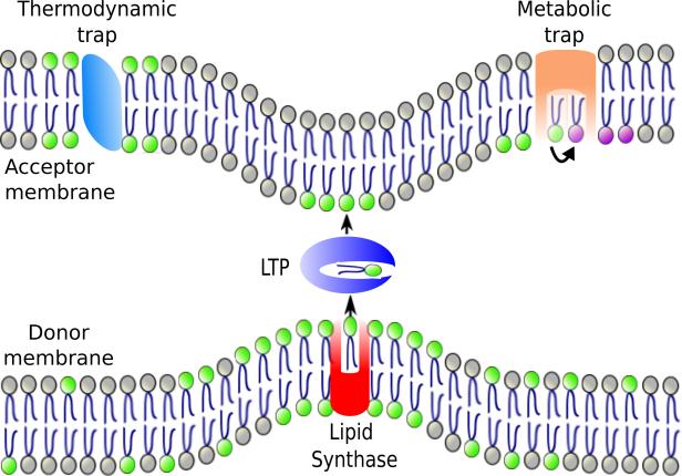
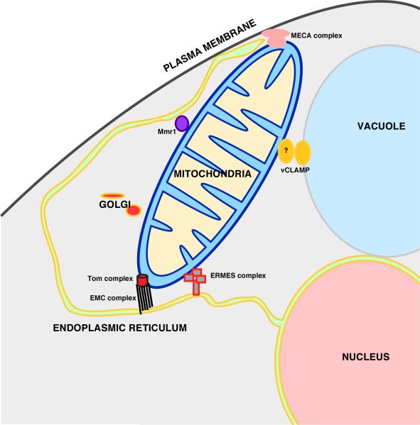
Maintaining the proper lipid composition of cellular membranes is critical for numerous cellular processes but mechanisms of membrane lipid homeostasis are not well understood. There is growing evidence that membrane contact sites (MCSs), regions where two organelles come in close proximity to one another, play major roles in the regulation of intracellular lipid composition and distribution. MCSs are thought to mediate the exchange of lipids and signals between organelles. In this review, we discuss how lipid exchange occurs at MCSs and evidence for roles of MCSs in regulating lipid synthesis and degradation. We also discuss how networks of organelles connected by MCSs may modulate cellular lipid homeostasis and help determine organelle lipid composition.


Figures

Figure 1
Figure 1. Model of directional lipid exchange at an MCS
A lipid synthase (red) localizes to an MCS where it produces a lipid (lipid with green headgroup). The enrichment of the lipid at MCS increases the likelihood that the lipid is transferred by an LTP at the MCSs. Alternatively, the synthase may hand the lipid directly to the LTP. Once the lipid reaches the second membrane it could be trapped there because of its affinity for a protein or lipid (thermodynamic trap) or by being modified (converted to a purple headgroup lipid; metabolic trap).
Figure 2
Figure 2. Lipid movement at the ER-mitochondria contacts
Lipids that move from ER to mitochondria (green arrows) or from mitochondria to the ER (red arrows) are shown. Black arrows indicate conversion of one lipid to another. PA = phosphatidic acid; CDP-DAG = cytidine diphosphate diacylglycerol; PS = phosphatidylserine; PE = phosphatidylethanolamine; PC = phosphatidylcholine; CL = cardiolipin.
Figure 3
Figure 3. Mitochondria centered interorganelle network in yeast
MCSs formed between mitochondria and other organelles in yeast and the protein complexes found at these MCSs. Some of these complexes are described in the text. The other complexes are thought to tether mitochondria to various organelles but have not yet shown to mediate lipid exchange. Tthe mitochondria ER cortex anchored (MECA) complex tethers mitochondrial, plasma membrane, and the ER [6]. A complex containing Mmr1 and other unknown proteins tethers mitochondria to the ER [45].

References

    1. Shevchenko A, Simons K. Lipidomics: coming to grips with lipid diversity. Nat Rev Mol Cell Biol. 2010:11593–598. - PubMed
    1. van Meer G, Voelker DR, Feigenson GW. Membrane lipids: where they are and how they behave. Nat Rev Mol Cell Biol. 2008:9112–124. - PMC - PubMed
    1. Helle SC, Kanfer G, Kolar K, Lang A, Michel AH, Kornmann B. Organization and function of membrane contact sites. Biochim Biophys Acta. 2013:18332526–2541. - PubMed
    1. Prinz WA. Bridging the gap: membrane contact sites in signaling, metabolism, and organelle dynamics. J Cell Biol. 2014:205759–769. - PMC - PubMed
    1. Knoblach B, Sun X, Coquelle N, Fagarasanu A, Poirier RL, Rachubinski RA. An ER-peroxisome tether exerts peroxisome population control in yeast. EMBO J. 2013:322439–2453. - PMC - PubMed

Publication types

LinkOut - more resources