

Assessment of white matter microstructure in stroke patients using NODDI
- PMID: 25570065
- PMCID: PMC4440535
- DOI: 10.1109/EMBC.2014.6943697
Assessment of white matter microstructure in stroke patients using NODDI
Abstract
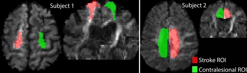
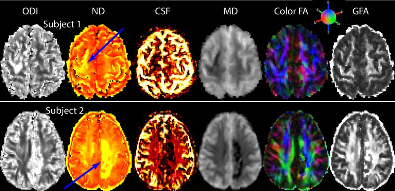
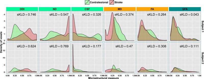
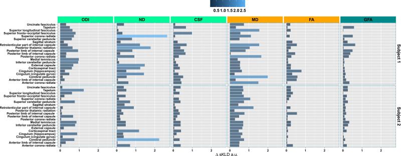
Diffusion weighted imaging (DWI) is widely used to study changes in white matter following stroke. In various studies employing diffusion tensor imaging (DTI) and high angular resolution diffusion imaging (HARDI) modalities, it has been shown that fractional anisotropy (FA), mean diffusivity (MD), and generalized FA (GFA) can be used as measures of white matter tract integrity in stroke patients. However, these measures may be non-specific, as they do not directly delineate changes in tissue microstructure. Multi-compartment models overcome this limitation by modeling DWI data using a set of indices that are directly related to white matter microstructure. One of these models which is gaining popularity, is neurite orientation dispersion and density imaging (NODDI). This model uses conventional single or multi-shell HARDI data to describe fiber orientation dispersion as well as densities of different tissue types in the imaging voxel. In this paper, we apply for the first time the NODDI model to 4-shell HARDI stroke data. By computing NODDI indices over the entire brain in two stroke patients, and comparing tissue regions in ipsilesional and contralesional hemispheres, we demonstrate that NODDI modeling provides specific information on tissue microstructural changes. We also introduce an information theoretic analysis framework to investigate the non-local effects of stroke in the white matter. Our initial results suggest that the NODDI indices might be more specific markers of white matter reorganization following stroke than other measures previously used in studies of stroke recovery.
Figures

References
-
- Daducci A, Canales-Rodriguez EJ, Descoteaux M, et al. Quantitative comparison of reconstruction methods for intra-voxel fiber recovery from diffusion MRI. Medical Imaging, IEEE Transactions on. 2014 Feb;33(2):384–399. - PubMed
-
- Granziera C, Daducci A, Meskaldji D, et al. A new early and automated MRI-based predictor of motor improvement after stroke. Neurology. 2012 Jul;79(1):39–46. - PubMed
-
- Granziera C, Daducci A, et al. Diffusion Spectrum Imaging after stroke shows structural changes in the contra-lateral motor network correlating with functional recovery. ISMRM. 2011:4199.
Publication types
MeSH terms
Grants and funding
LinkOut - more resources
Full Text Sources
Other Literature Sources
Medical