

Implementation of the excitatory entorhinal-dentate-CA3 topography in a large-scale computational model of the rat hippocampus
- PMID: 25571504
- PMCID: PMC4494103
- DOI: 10.1109/EMBC.2014.6945136
Implementation of the excitatory entorhinal-dentate-CA3 topography in a large-scale computational model of the rat hippocampus
Abstract
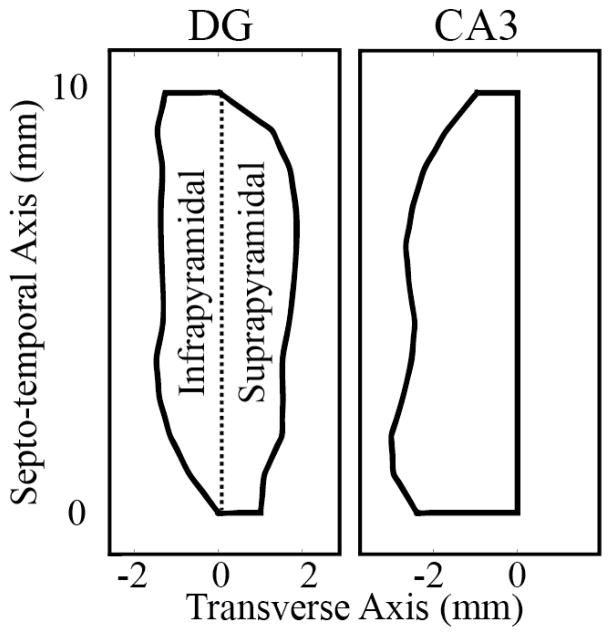
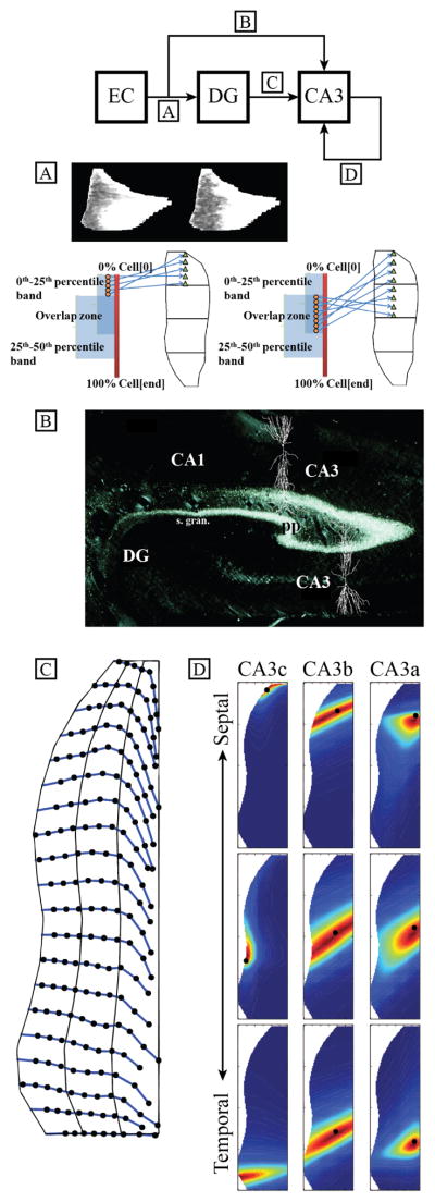
The topography, or the anatomical connectivity, of the excitatory entorhinal-dentate-CA3 circuit of the rat hippocampus has been implemented for a large-scale, biologically realistic, computational model of the rat hippocampus. The implementation thus far covers only the excitatory synapses for the principal neurons in the hippocampal subregions. Starting from layer II of the entorhinal cortex, the projection of their perforant path axons has been mapped across the full extent of the dentate gyrus as well as to the CA3. The mossy fiber axon trajectories from the dentate granule cells to the CA3 pyramidal cells have been derived, incorporating the transverse route the fibers take through the CA3c and CA3b and the septo-temporal turn in the CA3a. The extensive arborization of the CA3 pyramidal axons have been modeled using 2-D, skewed Gaussian distributions which have been parametrized to exhibit the differences that exist among the CA3a, CA3b, and CA3c auto-associational projections. Using the limited samples available from the literature, key parameters for each projection have been interpolated as a function of transverse and/or septo-temporal position in order to create a more complete representation of the topography.
Figures

Similar articles
-
Topography-dependent spatio-temporal correlations in the entorhinal-dentate-CA3 circuit in a large-scale computational model of the Rat Hippocampus.Annu Int Conf IEEE Eng Med Biol Soc. 2015;2015:3965-8. doi: 10.1109/EMBC.2015.7319262. Annu Int Conf IEEE Eng Med Biol Soc. 2015. PMID: 26737162 Free PMC article.
-
The CA3 "backprojection" to the dentate gyrus.Prog Brain Res. 2007;163:627-37. doi: 10.1016/S0079-6123(07)63034-9. Prog Brain Res. 2007. PMID: 17765742 Free PMC article. Review.
-
Convergence of entorhinal and CA3 inputs onto pyramidal neurons and interneurons in hippocampal area CA1--an anatomical study in the rat.Hippocampus. 2008;18(3):266-80. doi: 10.1002/hipo.20385. Hippocampus. 2008. PMID: 18000818
-
Functional interconnections between CA3 and the dentate gyrus revealed by current source density analysis.Hippocampus. 1998;8(3):217-30. doi: 10.1002/(SICI)1098-1063(1998)8:3<217::AID-HIPO5>3.0.CO;2-G. Hippocampus. 1998. PMID: 9662137
-
Ultrastructure and synaptic connectivity of cell types in the adult rat dentate gyrus.Prog Brain Res. 2007;163:155-66. doi: 10.1016/S0079-6123(07)63009-X. Prog Brain Res. 2007. PMID: 17765717 Review.
Cited by
-
Dentate gyrus granule cell activation following extracellular electrical stimulation: a multi-scale computational model to guide hippocampal neurostimulation strategies.Front Comput Neurosci. 2025 Aug 1;19:1638002. doi: 10.3389/fncom.2025.1638002. eCollection 2025. Front Comput Neurosci. 2025. PMID: 40822710 Free PMC article.
-
Network Activity Due to Topographic Organization of Schaffer Collaterals in a Large-Scale Model of Rat CA1.Annu Int Conf IEEE Eng Med Biol Soc. 2019 Jul;2019:2977-2980. doi: 10.1109/EMBC.2019.8856799. Annu Int Conf IEEE Eng Med Biol Soc. 2019. PMID: 31946514 Free PMC article.
-
Topographic Organization of Correlation Along the Longitudinal and Transverse Axes in Rat Hippocampal CA3 Due to Excitatory Afferents.Front Comput Neurosci. 2020 Nov 20;14:588881. doi: 10.3389/fncom.2020.588881. eCollection 2020. Front Comput Neurosci. 2020. PMID: 33328947 Free PMC article.
-
Volterra representation enables modeling of complex synaptic nonlinear dynamics in large-scale simulations.Front Comput Neurosci. 2015 Sep 17;9:112. doi: 10.3389/fncom.2015.00112. eCollection 2015. Front Comput Neurosci. 2015. PMID: 26441622 Free PMC article.
-
Topography-dependent spatio-temporal correlations in the entorhinal-dentate-CA3 circuit in a large-scale computational model of the Rat Hippocampus.Annu Int Conf IEEE Eng Med Biol Soc. 2015;2015:3965-8. doi: 10.1109/EMBC.2015.7319262. Annu Int Conf IEEE Eng Med Biol Soc. 2015. PMID: 26737162 Free PMC article.
References
-
- Gaarskjaer FB. Organization of the mossy fiber system of the rat studied in extended hippocampi. I. Terminal area related to number of granule and pyramidal cells. Journal of comparative neurology. 1978 Mar;178(1):49–72. - PubMed
-
- Ishizuka N, Weber J, Amaral DG. Organization of intrahippocampal projections originating from CA3 pyramidal cells in rat. Journal of comparative neurology. 1990 May;295(4):580–623. - PubMed
-
- Dolorfo CL, Amaral DG. Entorhinal cortex of the rat: topographic organization of the cells of origin of the perforant path projection to the dentate gyrus. Journal of Comparative Neurology. 1998 Aug;398(1):25–48. - PubMed
-
- Tamamaki N, Nojyo Y. Projection of the entorhinal layer II neurons in the rat as revealed by intracellular pressure-injection of neurobiotin. Hippocampus. 1993 Oct;3(4):471–80. - PubMed
Publication types
MeSH terms
Grants and funding
LinkOut - more resources
Full Text Sources
Other Literature Sources
Miscellaneous