. 2015 May;11(5):3308-16.
doi: 10.3892/mmr.2015.3146. Epub 2015 Jan 7.

Effect of Melilotus suaveolens extract on pulmonary microvascular permeability by downregulating vascular endothelial growth factor expression in rats with sepsis

Affiliations

Effect of Melilotus suaveolens extract on pulmonary microvascular permeability by downregulating vascular endothelial growth factor expression in rats with sepsis

Ming-Wei Liu et al. Mol Med Rep. 2015 May.

Abstract

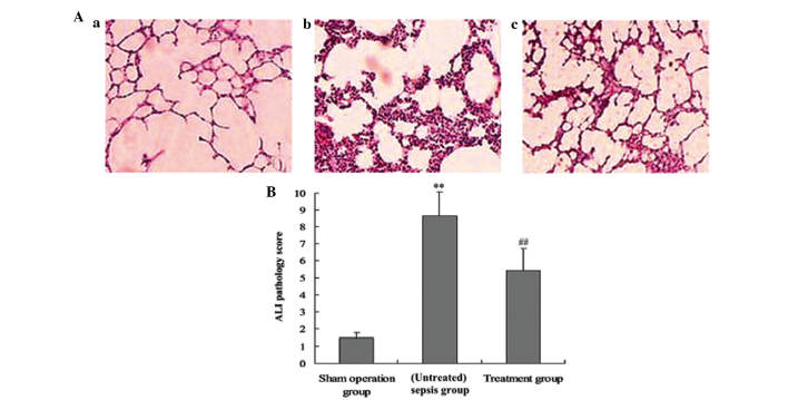
A typical indicator of sepsis is the development of progressive subcutaneous and body‑cavity edema, which is caused by the breakdown of endothelial barrier function, leading to a marked increase in vascular permeability. Microvascular leakage predisposes to microvascular thrombosis, breakdown of microcirculatory flow and organ failure, which are common events preceding mortality in patients with severe sepsis. Melilotus suaveolens (M. suaveolens) is a Traditional Tibetan Medicine. Previous pharmacological studies have demonstrated that an ethanolic extract of M. suaveolens has powerful anti‑inflammatory activity and leads to an improvement in capillary permeability. However, the mechanisms underlying its pharmacological activity remain elusive. The present study aimed to assess the impact of M. suaveolens extract tablets on pulmonary vascular permeability, and their effect on regulating lung inflammation and the expression of vascular endothelial growth factor (VEGF) in the lung tissue of rats with sepsis. A cecal ligation and puncture (CLP) sepsis model was established for both the control and treatment groups. ~2 h prior to surgery, 25 mg/kg of M. suaveolens extract tablet was administered to the treatment group. Polymerase chain reaction and western blot analyses were used to assess the expression of nuclear factor (NF)‑κB and VEGF in the lung tissue, and ELISA was applied to detect changes in serum tumor necrosis factor‑α as well as interleukins (IL) ‑1, ‑4, ‑6, and ‑10. The lung permeability, wet/dry weight ratio and lung pathology were determined. The results demonstrated that in the lung tissue of CLP‑rats with sepsis, M. suaveolens extract inhibited the expression of NF‑κB, reduced the inflammatory response and blocked the expression of VEGF, and thus significantly decreased lung microvascular permeability. The effects of M. Suaveolens extract may be of potential use in the treatment of CLP‑mediated lung microvascular permeability.


Figures

Figure 1
Figure 1
M. suaveolens extract blocked the expression of VEGF and NF-κB mRNA in lung tissue. Groups of mice were challenged with cecal ligation and puncture and treated with M. suaveolens extract 24 h later. The left lung tissues were homogenized and total RNA was extracted using TRIzol reagent and assayed by quantitative polymerase chain reaction. (A) Representative gels assessing (a) VEGF and (b) NF-κB levels are demonstrated. Lane 1, marker; lane 2, normal control group; lane 3, sham operation group; lane 4, (untreated) sepsis group and lane 5, treatment group. (B) Statistical summary of the densitometric analysis of VEGF and NF-κB mRNA expression in rats from the four groups; (a) VEGF mRNA and (b) NF-κB mRNA. Data are represented as the mean ± standard deviation of one experiment consisting of three replicates. **P<0.01 vs. sham operation and normal control groups; ##P<0.01 vs. (untreated) sepsis group. VEGF, vascular endothelial growth factor; NF-κB, nuclear factor kappa B.
Figure 2
Figure 2
Effect of M. suaveolens extract on the expression of VEGF and NF-κB65 protein in lung tissue. Groups of mice were challenged with cecal ligation and puncture and treated with M. suaveolens extract 24 h later. The expression of VEGF, NF-κβ65 and GAPDH was detected by western blotting using specific antibodies. GAPDH protein was used an internal control. (A) Representative western blot analysis demonstrated the levels of VEGF and NF-κB65 protein expression in rats from the four groups; (a) normal control group; (b) sham operation group; (c) (untreated) sepsis group and (d) treatment group. (B) Quantification of the blots by densitometric analysis of (a) VEGF and (b) NF-κB65 protein expression in rats from the four groups. Data are presented as the mean ± standard deviation of one experiment consisting of three replicates. **P<0.01 vs. the sham operation group and normal control group; #P<0.05 vs. (untreated) sepsis group. VEGF, vascular endothelial growth factor; NF-κB, nuclear factor kappa B.
Figure 3
Figure 3
Effect of M. suaveolens extract on the protein expression of VEGF and NF-κB65 in rat lungs 24 h following cecal ligation and puncture-induced acute lung injury. Immunostaining was performed on lung sections following antigen retrieval using Retrievagen. (A) Representative immunostaining revealed VEGF and NF-κβ65-positive expression in rats from the four groups: (a–c) Expression of positive VEGF in the (a) sham operation group; (b) control group; (c) treatment group); (d–f) Expression of positive NF-κB65 in the (d) sham operation group; (e) (untreated) sepsis group; (f) treatment group (magnification, ×200). (B) Quantification of the images by densitometric analysis of (a) VEGF and (b) NF-κB-positive protein expression in rats from four groups. All values are expressed as the mean ± standard deviation. **P<0.01 vs. the sham operation group; ##P<0.01 vs. (untreated) sepsis group. VEGF, vascular endothelial growth factor; NF-κB, nuclear factor kappa B.
Figure 4
Figure 4
Effect of M. suaveolens extract on plasma levels of VEGF, TNF-α, IL-6, IL-1β, IL-4 and IL-10 levels in plasma. Groups of mice were challenged with cecal ligation and puncture, and treated with M. suaveolens extract 24 h later. (A) VEGF, (B) TNF-α, (C) IL-6, (D) IL-1β, (E) IL-10 and (F) IL-4 levels in plasma were determined by ELISA. Data are presented as the mean ± standard deviation of one experiment consisting of three replicates. *P<0.05, **P<0.01 vs. the sham operation group and normal control group; #P<0.05, ##P<0.01 vs. (untreated) sepsis group. VEGF, vascular endothelial growth factor; NF-κB, nuclear factor kappa B; TNF-α, tumor necrosis factor-α; IL, interleukin.
Figure 5
Figure 5
Administration of M. suaveolens extract attenuated lipopolysaccharide-induced pulmonary inflammation. Groups of mice were challenged with cecal ligation and puncture, and treated with M. suaveolens extract 24 h later. (A) VEGF, (B) TNF-α, (C) IL-6, (D) IL-1β, (E) IL-10 and (F) IL-4 levels in bronchoalveolar lavage were determined by ELISA. Data are presented as the mean ± standard deviation of one experiment consisting of three replicates. **P<0.01 vs. the sham operation group and normal control group; #P<0.05, ##P<0.01 vs. (untreated) sepsis group. VEGF, vascular endothelial growth factor; NF-κB, nuclear factor kappa B; TNF-α, tumor necrosis factor-α; IL, interleukin.
Figure 6
Figure 6
Administration of M. suaveolens extract reduces CLP-induced lung permeability. Rats were treated as indicated and (A) FITC-labeled albumin in the bronchoalveolar lavage fluid, (B) water content of lung tissue and (C) W/D lung weight ratio were determined 24 h following CLP challenge. Data are presented as the mean ± standard deviation of one experiment consisting of three replicates. *P<0.05, **P<0.01 vs. the sham operation and normal control groups; ##P<0.01 vs. (untreated) sepsis group. VEGF, vascular endothelial growth factor; NF-κB, nuclear factor kappa B; TNF-α, tumor necrosis factor-α; IL, interleukin; CLP, cecal ligation and puncture; W/D, wet/dry; FITC, fluorescein isothiocyanate.
Figure 7
Figure 7
Administration of M. suaveolens extract ameliorated histopathologal changes in the lung tissue of CLP-ALI rats. The groups of rats were treated as described above and histological evaluation of the therapeutic potential of M. suaveolens extract on CLP-induced lung injury was performed 24 h following CLP challenge. (A) Representative images of hematoxylin and eosin-stained lung sections from three experimental groups (magnification, ×400): (a) Sham operation group; (b) (untreated) sepsis group; and (c) treatment group. (B) Lung injury score. ALI pathology score are expressed as the mean ± standard deviation of one experiment consisting of three replicates. **P<0.01 vs. the sham operation group; ##P<0.01 vs. (untreated) sepsis group. CLP, cecal ligation and puncture; ALI, acute lung injury.

Similar articles

Cited by

References

    1. Park SJ, Pai KS, Kim JH, Shin JI. What dose of intravenous immunoglobulin should be administered in Kawasaki disease with suspected systemic capillary leak syndrome? Comment on: shock: an unusual presentation of Kawasaki disease (Eur J Pediatr 2011 Jul; 170(7):941–3) Eur J Pediatr. 2012;17:203–204. doi: 10.1007/s00431-011-1602-7. - DOI - PubMed
    1. Zhao L, Tao JY, Zhang SL, et al. N-butanol extract from Melilotus Suaveolens Ledeb affects pro- and anti-inflammatory cytokines and mediators. Evid Based Complement Alternat Med. 2010;7:97–106. doi: 10.1093/ecam/nem165. - DOI - PMC - PubMed
    1. van Meurs M, Castro P, Shapiro NI, et al. Adiponectin diminishes organ-specific microvascular endothelial cell activation associated with sepsis. Shock. 2012;37:392–398. doi: 10.1097/SHK.0b013e318248225e. - DOI - PubMed
    1. Ueda T, Takeyama Y, Yasuda T, et al. Vascular endothelial growth factor increases in serum and protects against the organ injuries in severe acute pancreatitis. J Surg Res. 2006;134:223–230. doi: 10.1016/j.jss.2005.12.032. - DOI - PubMed
    1. Mura M, dos Santos CC, Stewart D, Liu M. Vaseular endothelial growth factor and related moleeules in acute lung injury. J Appl Physiol 1985. 2004;97:1605–1167. doi: 10.1152/japplphysiol.00202.2004. - DOI - PubMed

MeSH terms