Cranial nerve palsies in childhood
- PMID: 25572578
- PMCID: PMC4330297
- DOI: 10.1038/eye.2014.292
Cranial nerve palsies in childhood
Abstract
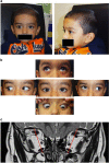
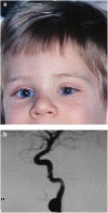
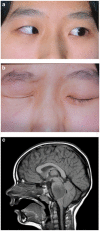
We review ocular motor cranial nerve palsies in childhood and highlight many of the features that differentiate these from their occurrence in adulthood. The clinical characteristics of cranial nerve palsies in childhood are affected by the child's impressive ability to repair and regenerate after injury. Thus, aberrant regeneration is very common after congenital III palsy; Duane syndrome, the result of early repair after congenital VI palsy, is invariably associated with retraction of the globe in adduction related to the innervation of the lateral rectus by the III nerve causing co-contraction in adduction. Clinical features that may be of concern in adulthood may not be relevant in childhood; whereas the presence of mydriasis in III palsy suggests a compressive aetiology in adults, this is not the case in children. However, the frequency of associated CNS abnormalities in III palsy and the risk of tumour in VI palsy can be indications for early neuroimaging depending on presenting features elicited through a careful history and clinical examination. The latter should include the neighbouring cranial nerves. We discuss the impact of our evolving knowledge of congenital cranial dysinnervation syndromes on this field.
Figures


References
-
- Holmes JM, Mutyala S, Maus TL, Grill R, Hodge DO, Gray DT. Pediatric third, fourth, and sixth nerve palsies: a population-based study. Am J Ophthalmol. 1999;127 (4:388–392. - PubMed
-
- Ng Ys, Lyons CJ. Oculomotor nerve palsy in childhood. Can J Ophthalmol. 2005;40 (5:645–653. - PubMed
-
- Schumacher-Feero LA, Yoo KW, Solari FM, Biglan AW. Third cranial nerve palsy in children. Am J Ophthalmol. 1999;128 (2:216–221. - PubMed
-
- Schatz NJ. Primary aberrant aculomotor regeneration. A sign of intracavernous meningioma. Arch Neurol. 1977;34 (1:29–32. - PubMed
-
- Loewenfeld IE, Thompson HS. Oculomotor paresis with cyclic spasms. A critical review of the literature and a new case. Surv Ophthalmol. 1975;20 (2:81–124. - PubMed
Publication types
MeSH terms
LinkOut - more resources
Full Text Sources
Other Literature Sources
Medical

