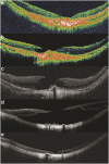
The development and evolution of full thickness macular hole in highly myopic eyes
- PMID: 25572579
- PMCID: PMC4366472
- DOI: 10.1038/eye.2014.312
The development and evolution of full thickness macular hole in highly myopic eyes
Abstract
Purpose: To evaluate the morphological changes before and after the formation of a full-thickness macular hole (MH) in highly myopic eyes.
Patients and methods: Retrospective observational case series. From 2006 to 2013, clinical records of patients with MH and high myopia who had optical coherence tomography (OCT) before the development of MH were reviewed. All patients had been followed for more than 1 year since MH formation to observe the morphological changes.
Results: Twenty-six eyes of 24 patients were enrolled. The initial OCT images could be classified into four types: (1) normal foveal depression with abnormal vitreo-retinal relationship (eight cases), (2) macular schisis without detachment (six cases), (3) macular schisis with concomitant/subsequent detachment (nine cases), and (4) macular atrophy with underlying/adjacent scar (three cases). After MH formation, one case in type 1 and one case in type 4 group developed retinal detachment (RD). In type 2 group, four cases developed RD at the same time of MH formation. The preexisting detachment in type 3 group extended in eight cases and improved in one case. Among all the cases, 14 eyes received vitrectomy and 7 eyes received gas injection. MH sealed in nine eyes after vitrectomy and four eyes by gas injection.
Conclusion: The study revealed four pathways of MH formation in highly myopic eyes. MH from macular schisis tended to be associated with detachment. However, the evolution and the results of surgical intervention were not always predictable.
Figures


Similar articles
-
Vitrectomy outcomes in eyes with high myopic macular hole without retinal detachment.Retina. 2012 Feb;32(2):275-80. doi: 10.1097/IAE.0b013e31821a8901. Retina. 2012. PMID: 21886021
-
Macular hole repair by vitrectomy and internal limiting membrane peeling in highly myopic eyes.Retina. 2014 Oct;34(10):2021-7. doi: 10.1097/IAE.0000000000000183. Retina. 2014. PMID: 24859476
-
Effect of 360° episcleral band as adjunctive to pars plana vitrectomy and silicone oil tamponade in the management of myopic macular hole retinal detachment.Retina. 2014 Apr;34(4):670-8. doi: 10.1097/IAE.0b013e3182a487ea. Retina. 2014. PMID: 24013260
-
Anatomical and visual outcomes in high myopic macular hole (HM-MH) without retinal detachment: a review.Graefes Arch Clin Exp Ophthalmol. 2014 Feb;252(2):191-9. doi: 10.1007/s00417-013-2555-5. Epub 2014 Jan 3. Graefes Arch Clin Exp Ophthalmol. 2014. PMID: 24384802 Review.
-
Surgical management of retinal detachment because of macular hole in highly myopic eyes.Retina. 2012 Oct;32(9):1704-18. doi: 10.1097/IAE.0b013e31826b671c. Retina. 2012. PMID: 23007668 Review.
Cited by
-
Myopic traction maculopathy in fovea-involved myopic chorioretinal atrophy.Eye (Lond). 2024 Dec;38(18):3586-3594. doi: 10.1038/s41433-024-03366-w. Epub 2024 Sep 23. Eye (Lond). 2024. PMID: 39313543
-
Risk factors for the development of idiopathic macular hole: a nationwide population-based cohort study.Sci Rep. 2022 Dec 16;12(1):21778. doi: 10.1038/s41598-022-25791-1. Sci Rep. 2022. PMID: 36526695 Free PMC article.
-
Fovea-sparing internal limiting membrane peeling with inverted flap technique versus standard internal limiting membrane peeling for symptomatic myopic foveoschisis.Sci Rep. 2024 Jan 30;14(1):2460. doi: 10.1038/s41598-024-53097-x. Sci Rep. 2024. PMID: 38291124 Free PMC article.
-
Macular assessment of preoperative optical coherence tomography in ageing Chinese undergoing routine cataract surgery.Sci Rep. 2018 Mar 23;8(1):5103. doi: 10.1038/s41598-018-22807-7. Sci Rep. 2018. PMID: 29572456 Free PMC article.
-
Development and validation of a deep learning system to classify aetiology and predict anatomical outcomes of macular hole.Br J Ophthalmol. 2023 Jan;107(1):109-115. doi: 10.1136/bjophthalmol-2021-318844. Epub 2021 Aug 4. Br J Ophthalmol. 2023. PMID: 34348922 Free PMC article.
References
-
- Yeh PT, Chen TC, Yang CH, Ho TC, Chen MS, Huang JS, et al. Formation of idiopathic macular hole—reappraisal. Graefes Arch Clin Exp Ophthalmol. 2010;248:793–798. - PubMed
-
- Miller JB, Yonekawa Y, Eliott D, Vavvas DG. A review of traumatic macular hole: diagnosis and treatment. Int Ophthalmol Clin. 2013;53 (4:59–67. - PubMed
-
- Yeh PT, Cheng CK, Chen MS, Yang CH, Yang CM. Macular hole in proliferative diabetic retinopathy with fibrovascular proliferation. Retina. 2009;29 (3:355–361. - PubMed
-
- Smiddy WE, Flynn HW., Jr Pathogenesis of macular holes and therapeutic implications. Am J Ophthalmol. 2004;137 (3:525–537. - PubMed
-
- Shih YH, Chiang TH, Hsiao CK, Chen CJ, Hung PT, Lin LK. Comparing Myopic Progression of Urban and Rural Taiwanese School children. Jpn J Ophthalmol. 2010;54:446–451. - PubMed
Publication types
MeSH terms
Substances
LinkOut - more resources
Full Text Sources
Other Literature Sources
Miscellaneous

