

The structural and compositional transition of the meniscal roots into the fibrocartilage of the menisci
- PMID: 25572636
- PMCID: PMC4304572
- DOI: 10.1111/joa.12265
The structural and compositional transition of the meniscal roots into the fibrocartilage of the menisci
Abstract
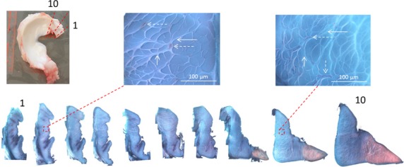
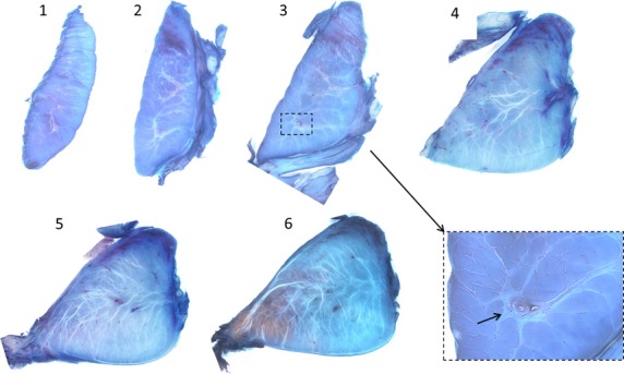
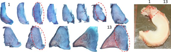
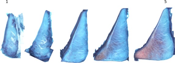
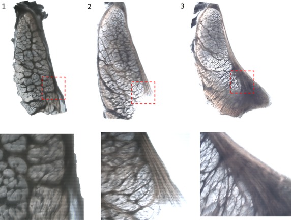
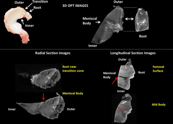
The meniscal roots, or insertional ligaments, firmly attach the menisci to tibial plateau. These strong attachments anchor the menisci and allow for the generation of hoop stress in the tissue. The meniscal roots have a ligament-like structure that transitions into the fibrocartilagenous structure of the meniscal body. The purpose of this study was to carry out a complete analysis of the structure and tissue organization from the body of the meniscus through the transition region and into the insertional roots. Serial sections were obtained from the meniscal roots into the meniscal body in fixed juvenile bovine menisci. Sections were stained for collagen and proteoglycans (PG) using fast green and safranin-o staining protocols. Unstained sections were imaged used a backlit stereo microscope. Optical projection tomography (OPT) was employed to evaluate the three-dimensional collagen architecture of the root-meniscus transition in lapine menisci. Tie-fibres were observed in the sections of the ligaments furthest from the bovine meniscal body. Blood vessels were observed to be surrounded by these tie-fibres and a PG-rich region within the ligaments. Near the tibial insertion, the roots contained large ligament-like collagen fascicles. In sections approaching the meniscus, there was an increase in tie-fibre size and density. Small tie-fibres extended into the ligament from the epiligamentous structure in the outermost sections of the meniscal roots, while large tie-fibre bundles were apparent at the meniscus transition. The staining pattern indicates that the root may continue into the outer portion of the meniscus where it then blends with the more fibrocartilage-like inner portions of the tissue. In unstained sections it was observed that the femoral side of the epiligamentous structure surrounding the root becomes more fibrous and thickens in the inferior inner portion of the posterior medial root. This thickening changes the shape of the root to more closely resemble the meniscus wedge shape. These observations support the concept of root continuity with the outer portion of the meniscus, thereby connecting with the hoop-like structure of the peripheral meniscus. OPT identified continuous collagen organization from the root into the meniscal body in longitudinal sections. In the radial direction, the morphology of the root continues into the meniscal body consistent with the serially sectioned bovine menisci. Blood vessels were prevalent on the periphery of the root. These blood vessels then arborized to cover the anterior femoral surface of the meniscus. This is the first study of the structural transition between the insertional ligaments (roots) and the fibrocartilagenous body of the menisci. These new structural details are important to understanding the meniscal load-bearing mechanism in the knee.
Keywords: Knee; Meniscal Roots; Meniscus; Optical Projection Tomography.
© 2015 Anatomical Society.
Figures
References
-
- Bedi A. Kelly N. Baad M, et al., editors. Dynamic contact mechanics of radial tears of the lateral meniscus: implications for treatment. Arthroscopy. 2012;28:372–381. - PubMed
-
- Gale D. Chaisson C. Totterman S, et al., editors. Meniscal subluxation: association with osteoarthritis and joint space narrowing. Osteoarthritis Cartilage. 1999;7:526–532. - PubMed
Publication types
MeSH terms
Substances
LinkOut - more resources
Full Text Sources
Other Literature Sources
Miscellaneous
