

Astrovirus VA1/HMO-C: an increasingly recognized neurotropic pathogen in immunocompromised patients
- PMID: 25572899
- PMCID: PMC4345817
- DOI: 10.1093/cid/ciu940
Astrovirus VA1/HMO-C: an increasingly recognized neurotropic pathogen in immunocompromised patients
Abstract
Background: An 18-month-old boy developed encephalopathy, for which extensive investigation failed to identify an etiology, 6 weeks after stem cell transplant. To exclude a potential infectious cause, we performed high-throughput RNA sequencing on brain biopsy.
Methods: RNA-Seq was performed on an Illumina Miseq, generating 20 million paired-end reads. Nonhost data were checked for similarity to known organisms using BLASTx. The full viral genome was sequenced by primer walking.
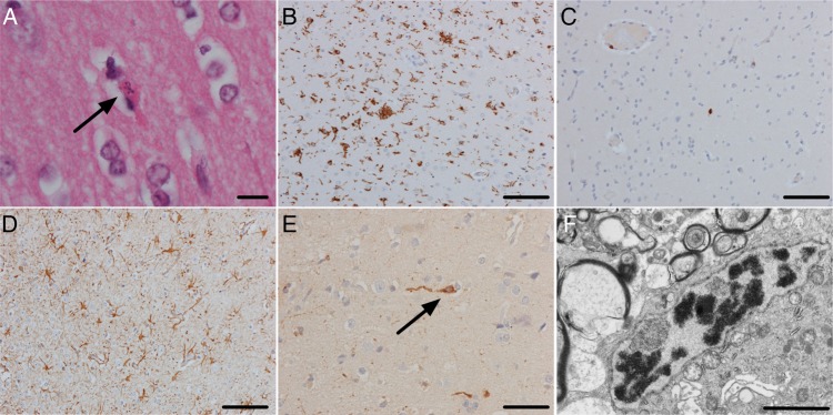
Results: We identified an astrovirus, HAstV-VA1/HMO-C-UK1(a), which was highly divergent from human astrovirus (HAstV 1-8) genotypes, but closely related to VA1/HMO-C astroviruses, including one recovered from a case of fatal encephalitis in an immunosuppressed child. The virus was detected in stool and serum, with highest levels in brain and cerebrospinal fluid (CSF). Immunohistochemistry of the brain biopsy showed positive neuronal staining. A survey of 680 stool and 349 CSF samples identified a related virus in the stool of another immunosuppressed child.
Conclusions: The discovery of HAstV-VA1/HMO-C-UK1(a) as the cause of encephalitis in this case provides further evidence that VA1/HMO-C viruses, unlike HAstV 1-8, are neuropathic, particularly in immunocompromised patients, and should be considered in the differential diagnosis of encephalopathy. With a turnaround from sample receipt to result of <1 week, we confirm that RNA-Seq presents a valuable diagnostic tool in unexplained encephalitis.
Keywords: RNASeq; astrovirus; deep sequencing; encephalopathy; pathogen discovery.
© The Author 2015. Published by Oxford University Press on behalf of the Infectious Diseases Society of America.
Figures
 ) and -UK1(b) (
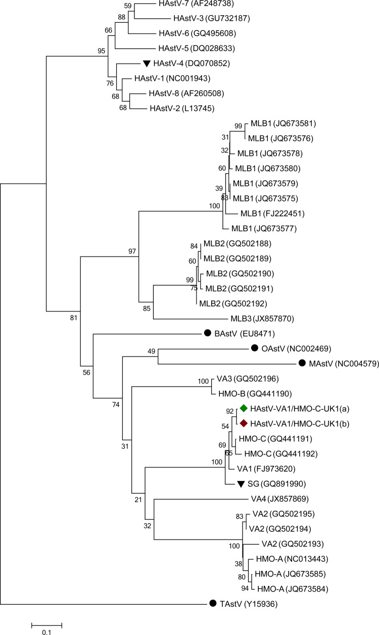
 ) to other astroviruses (AstVs) based on RNA-dependent RNA polymerase nucleotide sequences.
  indicates other AstV species that have been reported in patients with neurological disease.
  indicates sequences of animal origin, not human. Scale bar represents the number of base substitutions per site. Abbreviations: BAstV, bat astrovirus; MAstV, mink astrovirus; OAstV, ovine astrovirus; TAstV, turkey astrovirus.
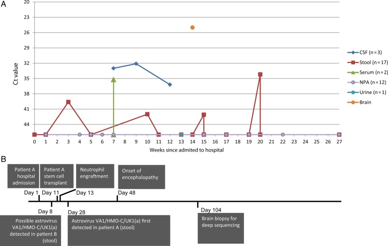
Comment in
-
Editorial commentary: Unbiased next-generation sequencing and new pathogen discovery: undeniable advantages and still-existing drawbacks.Clin Infect Dis. 2015 Mar 15;60(6):889-91. doi: 10.1093/cid/ciu913. Epub 2015 Jan 7. Clin Infect Dis. 2015. PMID: 25572900 No abstract available.
References
-
- Thompson C, Kneen R, Riordan A, Kelly D, Pollard AJ. Encephalitis in children. Arch Dis Child. 2012;97:150–61. - PubMed
-
- Koskiniemi M, Korppi M, Mustonen K, et al. Epidemiology of encephalitis in children. A prospective multicentre study. Eur J Pediatr. 1997;156:541–5. - PubMed
-
- Glaser CA, Honarmand S, Anderson LJ, et al. Beyond viruses: clinical profiles and etiologies associated with encephalitis. Clin Infect Dis. 2006;43:1565–77. - PubMed
-
- Johansson H, Bzhalava D, Ekstrom J, Hultin E, Dillner J, Forslund O. Metagenomic sequencing of “HPV-negative” condylomas detects novel putative HPV types. Virology. 2013;440:1–7. - PubMed
Publication types
MeSH terms
Substances
LinkOut - more resources
Full Text Sources
Other Literature Sources
