. 2015 May;36(5):863-70.
doi: 10.3174/ajnr.A4210. Epub 2015 Jan 8.

Neurovascular manifestations in hereditary hemorrhagic telangiectasia: imaging features and genotype-phenotype correlations

Collaborators, Affiliations

Neurovascular manifestations in hereditary hemorrhagic telangiectasia: imaging features and genotype-phenotype correlations

T Krings et al. AJNR Am J Neuroradiol. 2015 May.

Abstract

Background and purpose: Hereditary hemorrhagic telangiectasia is an autosomal dominant disease that presents in 10%-20% of patients with various brain vascular malformations. We aimed to report the radiologic features (phenotype) and the genotype-phenotype correlations of brain vascular malformations in hereditary hemorrhagic telangiectasia.

Materials and methods: Demographic, clinical, genotypic, and imaging information of 75 patients with hereditary hemorrhagic telangiectasia with brain arteriovenous malformations enrolled in the Brain Vascular Malformation Consortium from 2010 to 2012 were reviewed.

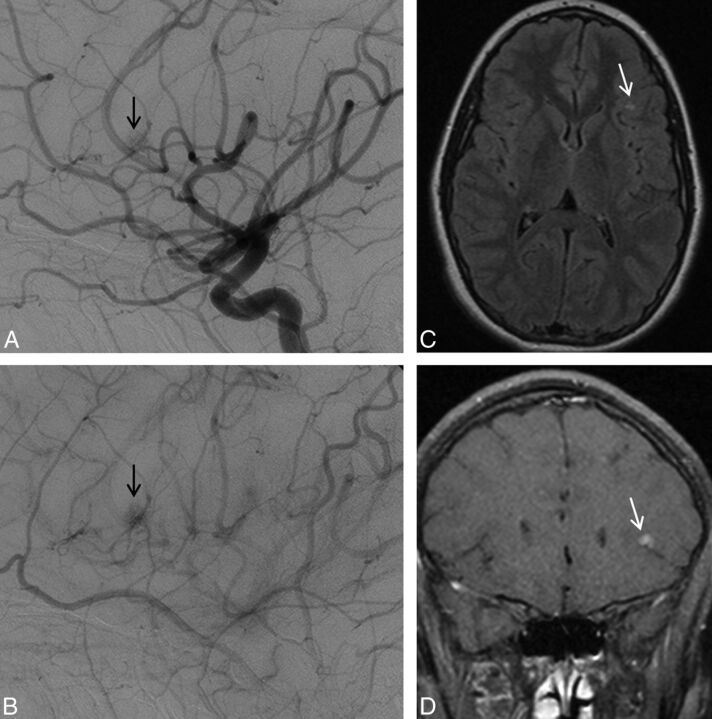
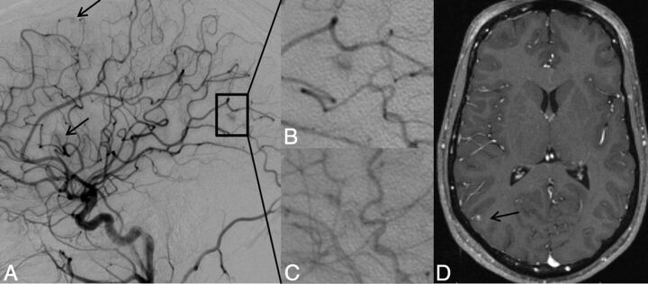
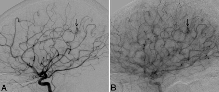
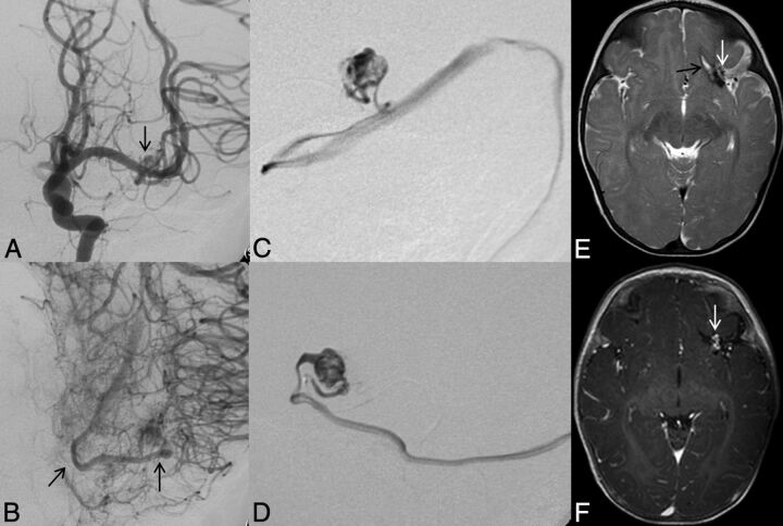
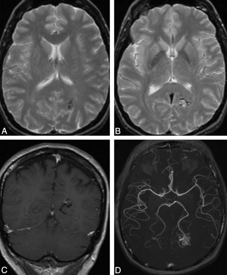
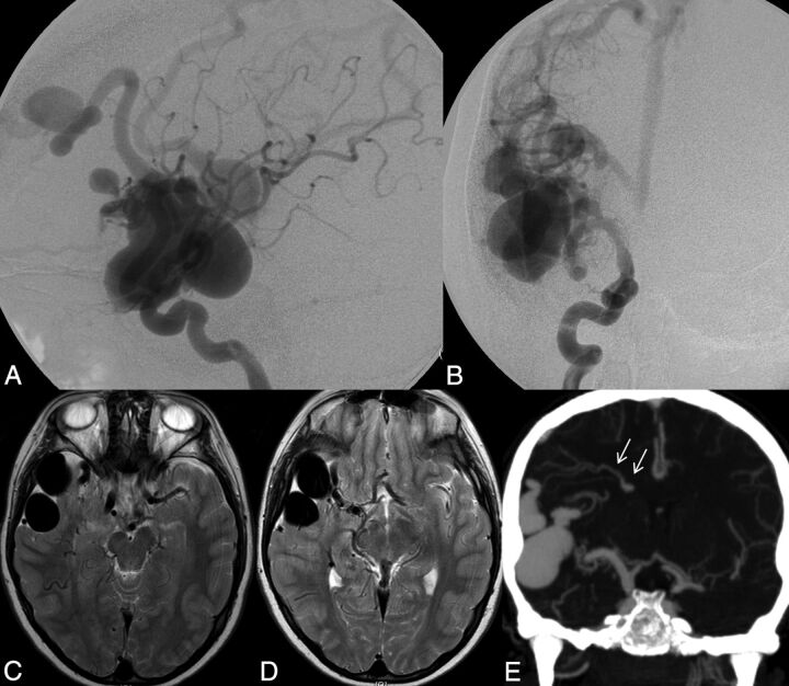
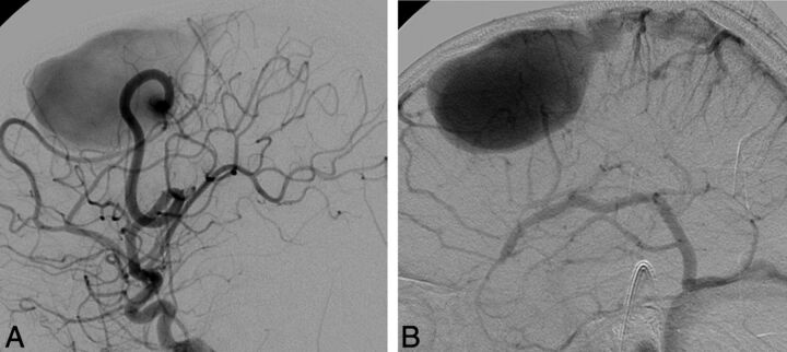
Results: Nonshunting, small, superficially located conglomerates of enhancing vessels without enlarged feeding arteries or draining veins called "capillary vascular malformations" were the most commonly observed lesion (46 of 75 patients; 61%), followed by shunting "nidus-type" brain AVMs that were typically located superficially with a low Spetzler-Martin Grade and a small size (32 of 75 patients; 43%). Direct high-flow fistulous arteriovenous shunts were present in 9 patients (12%). Other types of vascular malformations (dural AVF and developmental venous anomalies) were present in 1 patient each. Multiplicity of vascular malformations was seen in 33 cases (44%). No statistically significant correlation was observed between hereditary hemorrhagic telangiectasia gene mutation and lesion type or lesion multiplicity.

Conclusions: Depending on their imaging features, brain vascular malformations in hereditary hemorrhagic telangiectasia can be subdivided into brain AVF, nidus-type AVM, and capillary vascular malformations, with the latter being the most common phenotype in hereditary hemorrhagic telangiectasia. No genotype-phenotype correlation was observed among patients with this condition.


Figures

Fig 1.
Fig 1.
Left ICA angiogram, lateral view, depicts typical features of a capillary vascular malformation in the left frontal opercular region, with a subcentimeter vascular blush identified in the arterial (A) persisting into early venous phase (B, arrow), without evidence of arteriovenous shunting. On MR imaging, this appears as a focal region of hyperintensity on axial T2 FLAIR imaging (C, arrow), with fluffy enhancement on coronal gadolinium-enhanced T1-weighted imaging (D, arrow). No dilated arterial feeder or dilated draining vein is seen.
Fig 2.
Fig 2.
Lateral projection of right ICA angiogram (A–C) and axial T1-weighted gadolinium-enhanced MR imaging (D) demonstrate the typical imaging features of a capillary vascular malformation in a 17-year-old male patient with HHT. A focal subcentimeter blush (arrows) is identified on the arterial phase of a right carotid angiogram, in the frontal, temporal, and parietal regions, in keeping with multiple capillary telangiectasia. Magnified view of the right parietal lesion shows typical angiographic features, with vascular blush identified in the arterial (B) persisting into early venous phase (C); no arteriovenous shunting, dilated arterial feeder, or dilated draining vein are seen. MR imaging appearance of the right parietal lesion demonstrates the typical features of fluffy contrast enhancement in a superficial cortical/subcortical location, without abnormal vascular dilation (D, arrow).
Fig 3.
Fig 3.
Lateral view of a right ICA angiogram in a 43-year-old male patient with HHT demonstrates a capillary vascular malformation in the right anterior parietal region. A subcentimeter blush is seen in the arterial phase (A, arrow), which persists in the late arterial phase (B) without evidence of arteriovenous shunting.
Fig 4.
Fig 4.
Left ICA catheter angiogram (A and B), microcatheter injection (C and D), axial T2-weighted (E), and contrast-enhanced T1-weighted MR imaging (F) demonstrate imaging findings of a typical arteriovenous malformation. Anteroposterior projection of a left ICA injection demonstrates filling of the AVM nidus through an enlarged anterior temporal branch of the left MCA in the early arterial phase (A, arrow). There is arteriovenous shunting with early venous drainage through an enlarged left inferior temporal vein (B, arrows). Microcatheter injection in frontal (C) and lateral (D) projections demonstrates a typical glomerular well-defined compact nidus supplied by a single terminal arterial feeder with shunting into a dilated vein. MR imaging shows the superficial location of the AVM, with vascular flow voids seen on T2-weighted imaging (E, arrow) and enhancement on postgadolinium T1-weighted imaging (F, arrow). Evidence of previous hemorrhage related to AVM rupture, with a slit-like hematoma cavity in the left inferior frontal lobe, and surrounding hemosiderin staining (E, black arrow).
Fig 5.
Fig 5.
Typical MR imaging features of an arteriovenous malformation in a 50-year-old male patient with HHT. Axial T2-weighted imaging demonstrates a cluster of flow voids in the left occipital lobe, in keeping with a vascular nidus (A). A linear flow void of the dilated draining vein is seen inferior to the nidus on axial T2 (B) and coronal postcontrast T1-weighted imaging (C). Axial MRA MIP demonstrates a compact glomerular-type nidus in the left occipital lobe (D).
Fig 6.
Fig 6.
Right ICA catheter angiogram in lateral (A) and frontal (B) projections demonstrates 2 separate high-flow single-hole pial arteriovenous fistulas, 1 in the right frontal lobe and 1 in the right temporal lobe. Arterial supply to each fistula is through a single enlarged right MCA branch, which empties directly into a large venous pouch. Venous drainage is superficial, with both fistulas draining through enlarged cortical veins to the superior sagittal sinus. On axial T2-weighted MR imaging (C and D) venous pouches appear as flow voids and exert mass effect on the adjacent right temporal lobe. Evidence of venous congestion with venous reflux through dilated transdmedullary veins to the deep venous system is demonstrated on a coronal reconstruction of CTA (arrows).
Fig 7.
Fig 7.
Lateral view of a left ICA angiogram in an 8-year-old male patient with HHT. Angiography in early arterial (A) and venous (B) phases demonstrates a high-flow single-hole pial arteriovenous fistula in the left frontal lobe. Arterial supply is through a single enlarged prefrontal branch of the left MCA, which empties directly into a large venous pouch. Venous drainage is superficial through an enlarged cortical vein to the superior sagittal sinus.

References

    1. Bideau A, Plauchu H, Brunet G, et al. . Epidemiological investigation of Rendu-Osler disease in France: its geographical distribution and prevalence. Popul 1989;44:3–22 - PubMed
    1. Kjeldsen AD, Vase P, Green A. Hereditary haemorrhagic telangiectasia: a population-based study of prevalence and mortality in Danish patients. J Intern Med 1999;245:31–39 - PubMed
    1. Dakeishi M, Shioya T, Wada Y, et al. . Genetic epidemiology of hereditary hemorrhagic telangiectasia in a local community in the northern part of Japan. Hum Mutat 2002;19:140–48 - PubMed
    1. Fulbright RK, Chaloupka JC, Putman CM, et al. . MR of hereditary hemorrhagic telangiectasia: prevalence and spectrum of cerebrovascular malformations. AJNR Am J Neuroradiol 1998;19:477–84 - PMC - PubMed
    1. Matsubara S, Mandzia JL, ter Brugge K, et al. . Angiographic and clinical characteristics of patients with cerebral arteriovenous malformations associated with hereditary hemorrhagic telangiectasia. AJNR Am J Neuroradiol 2000;21:1016–20 - PMC - PubMed

Publication types

MeSH terms