

Intercellular adhesion molecule-1 expression in activated eosinophils is associated with mucosal remodeling in nasal polyps
- PMID: 25573100
- PMCID: PMC4368088
- DOI: 10.3892/mmr.2015.3174
Intercellular adhesion molecule-1 expression in activated eosinophils is associated with mucosal remodeling in nasal polyps
Abstract
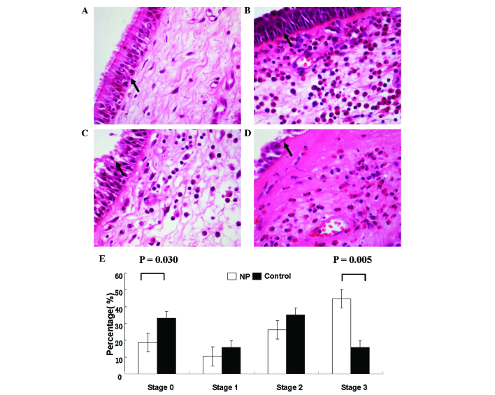
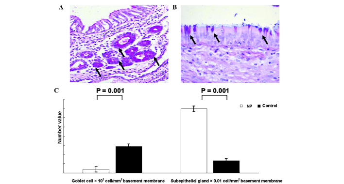
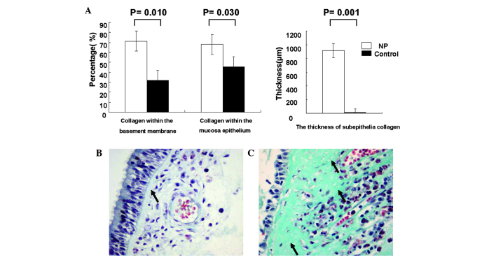
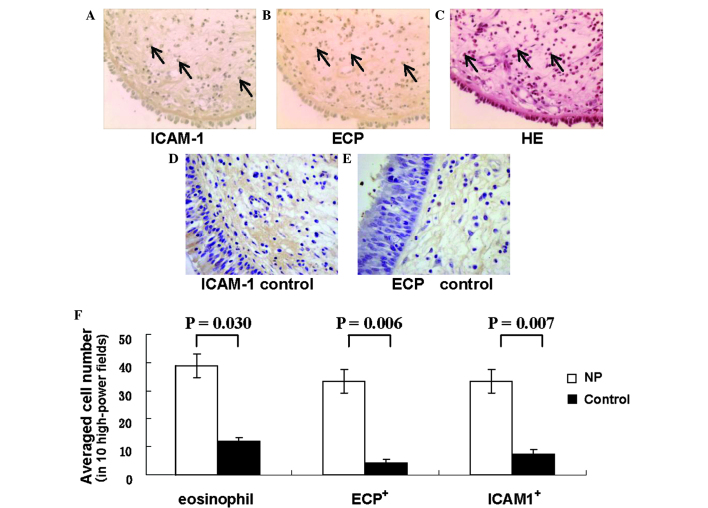
Nasal polyposis (NP) is characterized by chronic mucosal inflammation with infiltrating eosinophils. Eosinophil‑mediated tissue remodeling may be involved in NP pathogenesis; therefore, improved understanding of tissue remodeling may result the identification of novel pathways and therapeutic strategies. The present study aimed to investigate the pathological changes occurring during tissue remodeling in NP, in order to assess the role of intercellular adhesion molecule‑1 (ICAM‑1) in localized tissue remodeling and the potential association between ICAM‑1 expression and markers of eosinophil activation. A total of 28 eligible patients and 10 healthy controls participated in the current study. Nasal mucosal tissues of these subjects were retrospectively evaluated for mucosal remodeling using histopathological staining. ICAM‑1 and eosinophil cationic protein (ECP) expression levels were determined by immunohistochemical analysis. Compared with the healthy controls, all the specimens from NP patients presented substantial epithelial damage, skewed cellular distribution with a reduced density of goblet cells, an increased density of subepithelial gland and increased subepithelial collagen deposition. In addition, the NP specimens exhibited significantly higher eosinophil infiltration and ICAM‑1 expression compared with the controls. Positive correlations were observed between ICAM‑1 and ECP expression levels (P=0.010), as well as between extracellular collagen deposition and ICAM‑1 (P=0.010) and ECP (P=0.012) expression levels in the NP specimens, but not in the control specimens. Morphological evidence demonstrated eosinophil‑mediated tissue remodeling in NP tissues. ICAM‑1 expression in activated eosinophils was associated with NP remodeling, indicating the possibility that ICAM‑1 may regulate NP remodeling.
Figures
Similar articles
-
Eosinophils and mast cells: a comparison of nasal mucosa histology and cytology to markers in nasal discharge in patients with chronic sino-nasal diseases.Eur Arch Otorhinolaryngol. 2013 Sep;270(10):2667-76. doi: 10.1007/s00405-013-2395-2. Epub 2013 Feb 22. Eur Arch Otorhinolaryngol. 2013. PMID: 23430080
-
Measurement of inflammatory mediators of mast cells and eosinophils in native nasal lavage fluid in nasal polyposis.Int Arch Allergy Immunol. 2001 Jun;125(2):164-75. doi: 10.1159/000053811. Int Arch Allergy Immunol. 2001. PMID: 11435734
-
IL-25 as a novel therapeutic target in nasal polyps of patients with chronic rhinosinusitis.J Allergy Clin Immunol. 2015 Jun;135(6):1476-85.e7. doi: 10.1016/j.jaci.2015.01.003. Epub 2015 Feb 25. J Allergy Clin Immunol. 2015. PMID: 25725991
-
Pathogenesis of nasal polyps: an update.Curr Allergy Asthma Rep. 2005 Nov;5(6):463-71. doi: 10.1007/s11882-005-0027-7. Curr Allergy Asthma Rep. 2005. PMID: 16216171 Review.
-
The molecular biology of nasal polyposis.Curr Allergy Asthma Rep. 2001 May;1(3):262-7. doi: 10.1007/s11882-001-0017-3. Curr Allergy Asthma Rep. 2001. PMID: 11892044 Review.
Cited by
-
Wogonin attenuates nasal polyp formation by inducing eosinophil apoptosis through HIF-1α and survivin suppression.Sci Rep. 2018 Apr 18;8(1):6201. doi: 10.1038/s41598-018-24356-5. Sci Rep. 2018. PMID: 29670184 Free PMC article.
-
Exosomes: A Key Piece in Asthmatic Inflammation.Int J Mol Sci. 2021 Jan 19;22(2):963. doi: 10.3390/ijms22020963. Int J Mol Sci. 2021. PMID: 33478047 Free PMC article. Review.
References
-
- Kong H, Dong Z, Guo Y, Yang Z, Bu G. Intercellular adhesion molecule-1 and accumulation of eosinophils in nasal polyp tissue. Chin Med J (Engl) 1999;112:366–368. - PubMed
Publication types
MeSH terms
Substances
LinkOut - more resources
Full Text Sources
Other Literature Sources
Miscellaneous
