. 2015 Feb 16;21(8):3472-9.
doi: 10.1002/chem.201405535. Epub 2015 Jan 8.

Precise measurement of long-range heteronuclear coupling constants by a novel broadband proton-proton-decoupled CPMG-HSQMBC method

Affiliations

Precise measurement of long-range heteronuclear coupling constants by a novel broadband proton-proton-decoupled CPMG-HSQMBC method

István Timári et al. Chemistry. .

Abstract

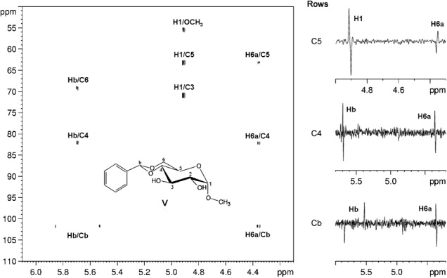
A broadband proton-proton-decoupled CPMG-HSQMBC method for the precise and direct measurement of long-range heteronuclear coupling constants is presented. The Zangger-Sterk-based homodecoupling scheme reported herein efficiently removes unwanted proton-proton splittings from the heteronuclear multiplets, so that the desired heteronuclear couplings can be determined simply by measuring frequency differences between singlet maxima in the resulting spectra. The proposed pseudo-1D/2D pulse sequences were tested on nucleotides, a metal complex incorporating P heterocycles, and diglycosyl (di)selenides, as well as on other carbohydrate derivatives, for the extraction of (n) J((1) H,(31) P), (n) J((1) H,(77) Se), and (n) J((1) H,(13) C) values, respectively.

Keywords: HSQMBC; NMR spectroscopy; heteronuclear coupling constants; proton-proton decoupling; structure elucidation.


Figures

Figure 1
Figure 1
Pulse sequence scheme of the broadband proton–proton-decoupled CPMG-HSQMBC experiment designed for the measurement of long-range heteronuclear coupling constants. Narrow and wide filled bars correspond to 90 and 180° pulses, respectively, with phase x unless indicated otherwise. The selective shaped proton pulse is shown as a half-ellipse. φ1 is incremented according to XY-16 cycles within the CPMG sequence; thus, n should ideally be adjusted to a multiple of 16. Other phases are φ2=y; φ3=x, −x; φ4=x, x, −x, −x; φ5=x, x, x, x, y, y, y, y; and φrec=x, −x, −x, x, −x, x, x, −x. Delays are set as follows: τ=120–150 μs, τa=1/(4*sw2), τb=1/(4*sw2)−4/sw. Coherence order selection and echo–antiecho phase-sensitive quadrature detection in the X dimension are achieved with gradient pulses G2 and G4 in the ratio 80:20.1 for 13C, 80:32.3846 for 31P and 80:15.257 for 77Se, respectively. Purging gradient pulses G1 and G3 are set to 19 and 10 % of maximum gradient strength (53 G cm−1). Coherence selection gradient pulses used in the extra proton–proton-decoupled dimension have G5=18 %. Sine-bell-shaped gradient pulses of 1 ms duration are utilized, followed by a recovery delay of 200 μs. The slice-selection gradient (G6) is adjusted for each molecule as reported in the legends to the respective figures.
Figure 2
Figure 2
Comparison of CPMG-HSQMBC spectra obtained for diglycosyl-selenide I, with (a) and without (b) broadband proton–proton decoupling. Measurement times were 5.9 h (a) and 45 min (b). The homonuclear decoupled pseudo-1D spectrum (a) was collected by using the sequence of Figure  1 with the incremented delay t1 replaced by a constant delay of 3 μs. Representative ZS-based PS 1H (c) and normal 1H NMR spectra (d) are also shown. All spectra in this figure were recorded with a spectral width of 6.0371 ppm. In the broadband proton–proton-decoupled spectra (a, c), an RSNOB selective 180° proton pulse[47] of duration 46.64 ms and bandwidth 50 Hz under a slice-selection gradient (G6) of 1 % of the maximum gradient strength was used. These spectra (a, c) were acquired with number of t2 increments (i.e., number of FID chunks)=32, duration of FID chunk=16.56 ms, number of complex data points of constructed FID in 1H dimension=3200, relaxation delay=2 s, number of scans=128 (a) and 4 (c). Spectrum b) was collected with number of complex data points=4096, relaxation delay=1.7 s, and number of scans=1024, by using the conventional CPMG-HSQMBC sequence.[17] The HSQMBC experiments (a, b) were recorded with 81.7 ms of heteronuclear coupling evolution during the initial CPMG-INEPT step.
Figure 3
Figure 3
Comparison of CPMG-HSQMBC spectra obtained for II, with (a) and without (b) broadband proton–proton decoupling. Measurement times were 1.5 h (a) and 10 min (b). The homonuclear decoupled pseudo-1D spectrum (a) was collected by using the sequence of Figure  1 with the incremented delay t1 replaced by a constant delay of 3 μs. Representative ZS-based PS 1H (c) and normal 1H NMR spectra (d) are also shown. Spectra a), c), and d) were recorded with spectral widths=6.0371 ppm. In the cases of the broadband proton–proton-decoupled spectra (a, c), an RSNOB selective 180° proton pulse[47] of duration 93.28 ms and bandwidth 25 Hz under a slice-selection gradient (G6) of 1 % of the maximum gradient strength was used. These spectra (a, c) were acquired with number of t2 increments (i.e., number of FID chunks)=32, duration of FID chunk=16.56 ms, number of complex data points of constructed FID in 1H dimension=3200, relaxation delay=1.7 s, number of scans=32 (a) and 8 (c). Spectrum b) was collected with spectral width=9.9774 ppm, number of complex data points=16 384, relaxation delay=1.7 s, and number of scans=128 by using the conventional CPMG-HSQMBC sequence.[17] The HSQMBC experiments (a, b) were recorded with 81.7 ms of heteronuclear coupling evolution during the initial CPMG-INEPT step.
Figure 4
Figure 4
Comparison of CPMG-HSQMBC spectra obtained for III, with (a) and without (b) broadband proton–proton decoupling. Measurement times were 3.3 h (a) and 13 min (b). The homonuclear decoupled pseudo-1D spectrum (a) was collected by using the sequence of Figure  1 with the incremented delay t1 replaced by a constant delay of 3 μs, and using an RSNOB selective 180° proton pulse[47] of duration 23.32 ms and bandwidth 100 Hz under a slice-selection gradient (G6) of 1.6 % of the maximum gradient strength. This spectrum (a) was recorded with spectral width=9.9774 ppm, number of t2 increments (i.e., number of FID chunks)=32, duration of FID chunk=20.04 ms, number of complex data points of constructed FID in 1H dimension=6400, relaxation delay=1.7 s, and number of scans=96. Spectrum b) was acquired with spectral width=9.9774 ppm, number of complex data points=8192, relaxation delay=2 s, and number of scans=256 by using the conventional 1D CPMG-HSQMBC sequence.[17] The HSQMBC experiments (a, b) were recorded with 52.7 ms of heteronuclear coupling evolution during the initial CPMG-INEPT step.
Figure 5
Figure 5
Representative broadband proton–proton-decoupled 2D CPMG-HSQMBC spectrum of diglycosyl diselenide IV. The extracted selenium traces shown next to the 2D spectrum nicely illustrate that the proposed method results in clean, pure absorptive antiphase doublets with splittings due solely to the desired multiple-bond heteronuclear couplings. The normal 1H spectrum can be seen above the 2D contour plot. The broadband proton–proton-decoupled CPMG-HSQMBC spectrum was recorded by using an RSNOB selective 180° proton pulse[47] of duration 46.64 ms and bandwidth 50 Hz under a slice-selection gradient (G6) of 0.5 % of the maximum gradient strength. The spectrum was acquired at 308 K in an experiment time of 18.3 h with spectral width in the 1H (77Se) dimension=9.9774 (140.0) ppm, number of t1 increments=32, number of t2 increments (i.e., number of FID chunks)=16, duration of FID chunk=16.56 ms, number of complex data points of constructed FID in 1H dimension=1600, relaxation delay=1.7 s, number of scans=48, and duration of long-range heteronuclear coupling evolution=81.7 ms.
Figure 6
Figure 6
Partial contour plot of the broadband proton–proton-decoupled 2D CPMG-HSQMBC spectrum of V. The extracted carbon traces shown next to the 2D spectrum show pure absorptive antiphase doublets with splittings due solely to the heteronuclear couplings. The spectrum was recorded with an RSNOB selective 180° proton pulse[47] of duration 46.64 ms and bandwidth 50 Hz under a slice-selection gradient (G6) of 1 % of the maximum gradient strength. The spectrum was acquired in an experiment time of 38.7 h with spectral width in the 1H (13C) dimension=6.0370 (80.0) ppm, number of t1 increments=200, number of t2 increments (i.e., number of FID chunks)=16, duration of FID chunk=21.12 ms, number of complex data points of constructed FID in 1H dimension=2048, relaxation delay=1.7 s, number of scans=16, and duration of long-range heteronuclear coupling evolution=74.4 ms.

Similar articles

Cited by

References

    1. Williamson RT, Buevich AV, Martin GE, Parella T. J. Org. Chem. 2014;79:3887–3894. - PubMed
    1. Matsumori N, Kaneno D, Murata M, Nakamura H, Tachibana K. J. Org. Chem. 1999;64:866–876. - PubMed
    1. Bifulco G, Dambruoso P, Gomez-Paloma L, Riccio R. Chem. Rev. 2007;107:3744–3779. - PubMed
    1. Marquez BL, Gerwick WH, Williamson RT. Magn. Reson. Chem. 2001;39:499–530.
    1. Parella T, Espinosa JF. Prog. Nucl. Magn. Reson. Spectrosc. 2013;73:17–55. - PubMed

Publication types

LinkOut - more resources