Review
. 2015 Jan 8;7(5):a016337.
doi: 10.1101/cshperspect.a016337.

Group 2 innate lymphoid cells in health and disease

Affiliations
Review

Group 2 innate lymphoid cells in health and disease

Brian S Kim et al. Cold Spring Harb Perspect Biol. .

Abstract

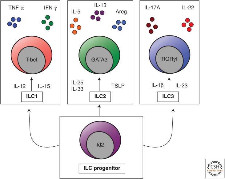
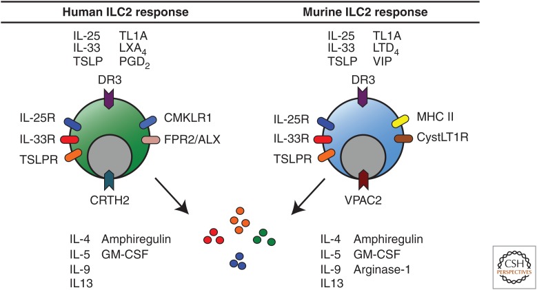
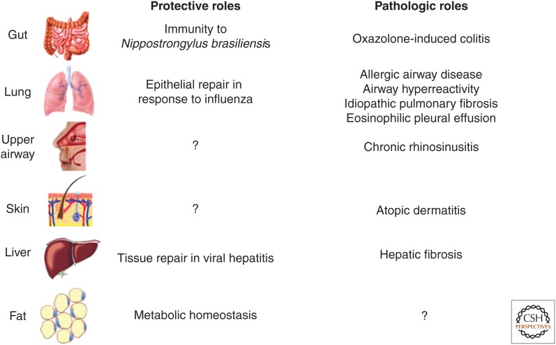
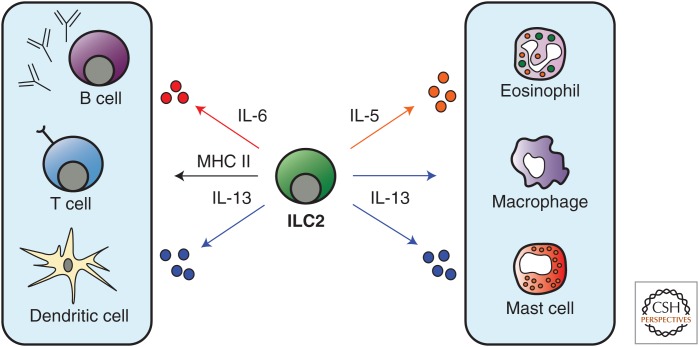
Group 2 innate lymphoid cells (ILC2s) play critical roles in anti-helminth immunity, airway epithelial repair, and metabolic homeostasis. Recently, these cells have also emerged as key players in the development of allergic inflammation at multiple barrier surfaces. ILC2s arise from common lymphoid progenitors in the bone marrow, are dependent on the transcription factors RORα, GATA3, and TCF-1, and produce the type 2 cytokines interleukin (IL)-4, IL-5, IL-9, and/or IL-13. The epithelial cell-derived cytokines IL-25, IL-33, and TSLP regulate the activation and effector functions of ILC2s, and recent studies suggest that their responsiveness to these cytokines and other factors may depend on their tissue environment. In this review, we focus on recent advances in our understanding of the various factors that regulate ILC2 function in the context of immunity, inflammation, and tissue repair across multiple organ systems.


Figures

Figure 1.
Figure 1.
The innate lymphoid cell family. Innate lymphoid cells (ILCs) are a heterogeneous family of innate immune cells that arise from a common Id2-dependent lymphoid progenitor in the bone marrow. Group 1 ILCs (ILC1s) respond to IL-12 and IL-15, express the transcription factor T-bet, and produce tumor necrosis factor α (TNF-α) and interferon γ (IFN-γ). Group 2 ILCs (ILC2s) respond to IL-25, IL-33, and thymic stromal lymphopoietin (TSLP), express the transcription factor GATA3, and produce IL-4, IL-5, IL-9, IL-13, and amphiregulin (Areg). Group 3 ILCs (ILC3s) respond to IL-23 and IL-1β, express the transcription factor RORγt, and produce IL-17A and IL-22.
Figure 2.
Figure 2.
Regulation of human and murine ILC2 responses. Human ILC2s are activated by IL-25, IL-33, TSLP, TL1A, and prostaglandin D2 (PGD2) and inhibited by lipoxin A4 (LXA4). Murine ILC2s are activated by IL-25, IL-33, TSLP, TL1A, leukotriene D4 (LTD4), and vasoactive intestinal peptide (VIP). Both human and murine ILC2s express the receptors to respond directly to these mediators and produce IL-4, IL-5, IL-9, IL-13, granulocyte macrophage colony–stimulating factor (GM-CSF), and amphiregulin (Areg). Murine ILC2s express arginase-1.
Figure 3.
Figure 3.
Protective versus pathogenic roles of ILC2s. Gut-associated ILC2s play an essential role in protective immunity to helminth parasites and can promote type 2 inflammatory colitis. Lung ILC2s have shown a protective role by mediating epithelial repair in response to influenza virus infection. Further, they have shown pathogenic roles by promoting allergic airway disease, airway hyperreactivity, and pulmonary fibrosis and have been implicated in eosinophilic pleural effusion. ILC2s in the upper airway have been implicated in chronic rhinosinusitis and found in the nasal poylps of patients. Skin ILC2s are highly enriched in atopic dermatitis (AD) lesions and promote AD-like disease in murine models. ILC2s in the liver have been shown to promote tissue repair in response to viral-induced injury as well as pathogenic fibrosis. ILC2s in white adipose tissue regulate metabolic homeostasis via cellular interactions with eosinophils and alternatively activated macrophages.
Figure 4.
Figure 4.
Effector functions of ILC2s. ILC2s in the lung have been proposed to promote adaptive CD4+ T-cell and B-cell responses via MHC class II–mediated antigen presentation and IL-6 production, respectively. Further, IL-13 has been shown to promote CD4+ T-cell responses indirectly via acting on dendritic cells. ILC2-derived IL-5 promotes eosinophil responses and IL-13 can regulate alternatively activated macrophage and mast cell functions.

Similar articles

Cited by

References

    1. Bando JK, Nussbaum JC, Liang HE, Locksley RM. 2013. Type 2 innate lymphoid cells constitutively express arginase-I in the naïve and inflamed lung. J Leukoc Biol 94: 877–884. - PMC - PubMed
    1. Barlow JL, Peel S, Fox J, Panova V, Hardman CS, Camelo A, Bucks C, Wu X, Kane CM, Neill DR, et al. 2013. IL-33 is more potent than IL-25 in provoking IL-13-producing nuocytes (type 2 innate lymphoid cells) and airway contraction. J Allergy Clin Immunol 132: 933–941. - PubMed
    1. Barnig C, Cernadas M, Dutile S, Liu X, Perrella MA, Kazani S, Wechsler ME, Israel E, Levy BD. 2013. Lipoxin A4 regulates natural killer cell and type 2 innate lymphoid cell activation in asthma. Sci Transl Med 5: 174ra126. - PMC - PubMed
    1. Bartemes KR, Iijima K, Kobayashi T, Kephart GM, McKenzie AN, Kita H. 2012. IL-33–responsive lineage– CD25+ CD44hi lymphoid cells mediate innate type 2 immunity and allergic inflammation in the lungs. J Immunol 188: 1503–1513. - PMC - PubMed
    1. Beamer CA, Girtsman TA, Seaver BP, Finsaas KJ, Migliaccio CT, Perry VK, Rottman JB, Smith DE, Holian A. 2012. IL-33 mediates multi-walled carbon nanotube (MWCNT)-induced airway hyper-reactivity via the mobilization of innate helper cells in the lung. Nanotoxicology 7: 1070–1081. - PMC - PubMed

Substances