. 2015 Jan 9;347(6218):170-5.
doi: 10.1126/science.1260580.

Gut microbiota. Antimicrobial peptide resistance mediates resilience of prominent gut commensals during inflammation

Affiliations

Gut microbiota. Antimicrobial peptide resistance mediates resilience of prominent gut commensals during inflammation

T W Cullen et al. Science. .

Abstract

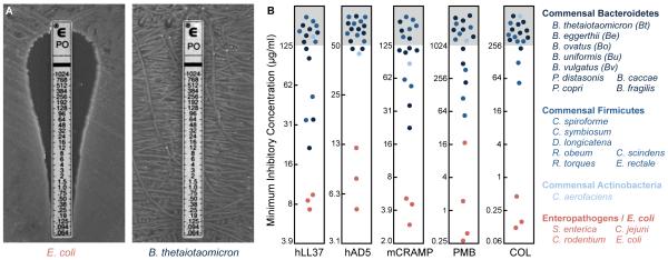
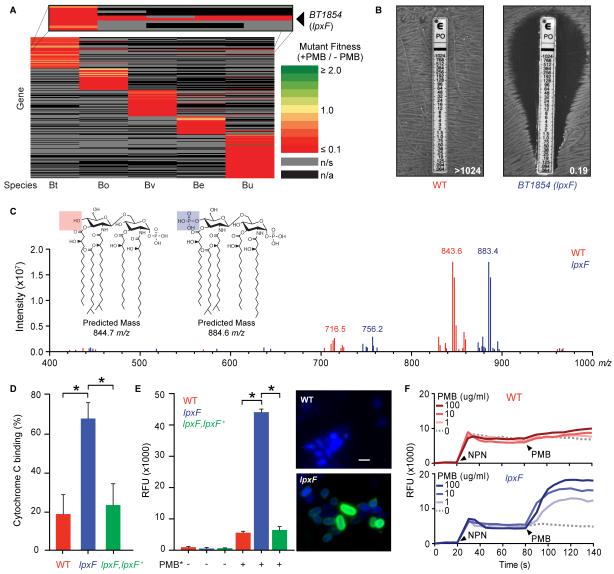
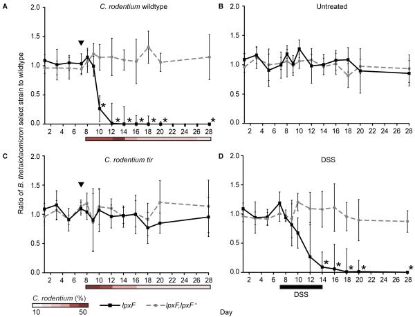
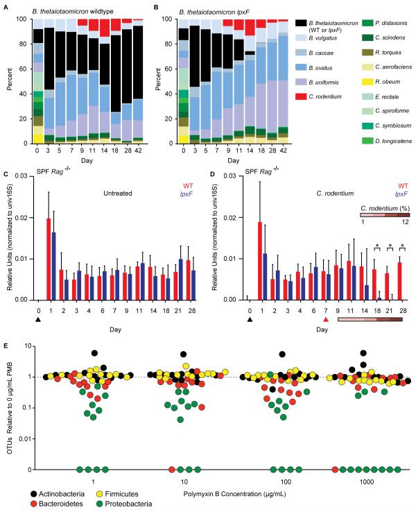
Resilience to host inflammation and other perturbations is a fundamental property of gut microbial communities, yet the underlying mechanisms are not well understood. We have found that human gut microbes from all dominant phyla are resistant to high levels of inflammation-associated antimicrobial peptides (AMPs) and have identified a mechanism for lipopolysaccharide (LPS) modification in the phylum Bacteroidetes that increases AMP resistance by four orders of magnitude. Bacteroides thetaiotaomicron mutants that fail to remove a single phosphate group from their LPS were displaced from the microbiota during inflammation triggered by pathogen infection. These findings establish a mechanism that determines the stability of prominent members of a healthy microbiota during perturbation.


Figures

Fig. 1
Fig. 1. Human gut commensals are highly resistant to cationic AMPs
(A) MIC of PMB against Escherichia coli and Bacteroides thetaiotaomicron. (B) MICs of human and mouse inflammation-induced AMPs and bacterial surrogates against prominent human gut commensal bacteria (blue) and enteropathogens (red). MICs of PMB and colistin were determined using E-test strips; others were determined using the microtiter broth dilution method. See also fig. S1 and table S1.
Fig. 2
Fig. 2. Lipid A dephosphorylation mediates AMP resistance in prominent human gut bacteria
(A) Heat map of the relative fitness of transposon mutant strains in the presence and absence of PMB. Columns indicate human gut bacterial species tested (abbreviations from Fig. 1B); rows indicate gene orthologs with insertions displaying a significantly altered fitness (q<0.05) in the presence of PMB in at least one species. n/s, not significantly altered; n/a, no ortholog. (B) BT1854 (LpxF) is necessary for PMB resistance in B. thetaiotaomicron. Representative MIC assessed using the E-test method is shown. (C) LpxF is required for dephosphorylation of lipid A. FT-ICR MS analysis of lipid A isolated from wildtype (red) and lpxF deletion mutant (blue) B. thetaiotaomicron revealed a shift of the predominant peak by ~40 m/z, consistent with the gain of a single phosphate group. Inset shows predicted structures and m/z values of the doubly deprotonated ([M-2H]2−) ions for monophosphorylated (red) and bis-phosphorylated (blue) penta-acylated lipid A. Minor peaks are consistent with tetra-acylated lipid A structures (fig. S2C-D). (D) Deletion of lpxF increases cationic cytochrome C binding to B. thetaiotaomicron, indicating altered surface charge. (E) Increased PMB-Oregon Green (PMB*) binding to lpxF deletion mutant cells measured by fluorescence quantification (left panel) and microscopy (right panels). Error bars represent standard deviation and asterisks indicate significance (p<0.01). Representative fluorescence microscopy images of bacterial cells incubated with PMB* (green) are shown as an overlay with 4′, 6′ diamino-2-phenylindole (DAPI) (blue). Scale bar indicates 2 μm. (F) LpxF protects the outer membrane from PMB perturbation. 1-N-phenylnaphthylamine (NPN) uptake profiles of select strains were measured followed by challenge with the indicated concentration of PMB. Arrowheads indicate the addition of NPN (20s) and PMB (80s). Readings were taken in 5s intervals. Each experiment was performed in triplicate with representative results shown. See figure S3 for complementation.
Fig. 3
Fig. 3. AMP resistance determines the resilience of a prominent human gut symbiont in gnotobiotic mice with colitis
Germfree mice (n=5/group) were colonized with wildtype, the lpxF deletion mutant (solid lines), and complemented (lpxF,lpxF+; dashed lines) B. thetaiotaomicron strains 7 days prior to initiation of inflammation by C. rodentium infection (A), no further treatment (B), infection with a non-inflammatory C. rodentium tir mutant (C), or exposure to 3% DSS ad libitum for 7 days (black bar) (D). Infection with C. rodentium strains is indicated by black arrowheads and relative C. rodentium abundances are reported as percent of total fecal DNA (shaded bars); error bars represent standard deviation and asterisks indicate significant (p<0.01) differences.
Fig. 4
Fig. 4. AMP resistance determines commensal resilience in the context of a human or murine gut microbiota
(A, B) Germfree mice (n=5/group) were colonized with 14 prominent human gut microbes, including B. thetaiotaomicron wildtype or lpxF deletion mutant, 7 days prior to infection with C. rodentium; community composition was monitored by species-specific quantitative polymerase chain reaction (qPCR) and reported as median percent of total. Uninfected controls are shown in fig. S6B. (C, D) Specific pathogen free Rag−/− mice (n=5/group) were gavaged (black arrowhead) with either B. thetaiotaomicron wildtype or lpxF deletion mutant; in (D), mice were infected with C. rodentium 7 days later (red arrowhead). Colonization levels were assessed by qPCR from fecal DNA. C. rodentium levels are reported percentage of total fecal DNA. Error bars indicate standard deviation and asterisks indicate significance (p<0.05). (E) AMP resistance is a general feature of human gut Bacteroidetes, Firmicutes, and Actinobacteria. Fecal samples from 12 unrelated, healthy human donors were cultured on varying PMB concentrations (x-axis) and the number of species-level phylotypes (OTUs) belonging to each phylum observed from each donor (points, colored by phylum) was normalized to the number observed in culture in the absence of PMB. A weighted (abundance) analysis provides similar results (fig. S8B).

Comment in

References

    1. Langhorst J, et al. Elevated human beta-defensin-2 levels indicate an activation of the innate immune system in patients with irritable bowel syndrome. Am. J. Gastroenterol. 2009;104:404–410. - PubMed
    1. Hooper LV, Littman DR, Macpherson AJ. Interactions between the microbiota and the immune system. Science. 2012;336:1268–1273. - PMC - PubMed
    1. Faith JJ, et al. The long-term stability of the human gut microbiota. Science. 2013;341:44–52. - PMC - PubMed
    1. Vaara M. Agents that increase the permeability of the outer membrane. Microbiol. Rev. 1992;56:395–411. - PMC - PubMed
    1. Qin J, et al. A human gut microbial gene catalogue established by metagenomic sequencing. Nature. 2010;464:59–65. - PMC - PubMed

Publication types

MeSH terms