. 2015 Apr 15;24(8):2375-89.
doi: 10.1093/hmg/ddv004. Epub 2015 Jan 7.

MATR3 disruption in human and mouse associated with bicuspid aortic valve, aortic coarctation and patent ductus arteriosus

Affiliations

MATR3 disruption in human and mouse associated with bicuspid aortic valve, aortic coarctation and patent ductus arteriosus

Fabiola Quintero-Rivera et al. Hum Mol Genet. .

Abstract

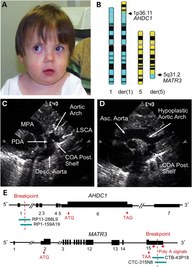
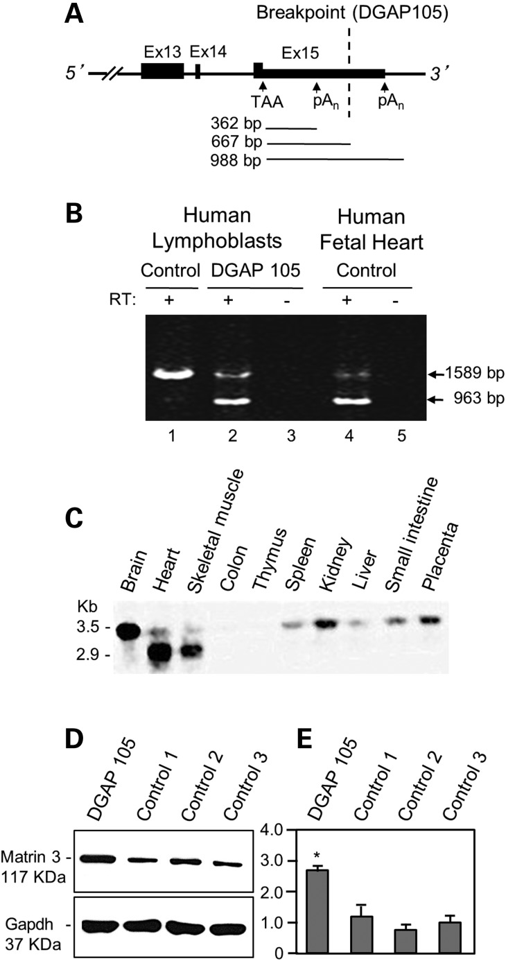
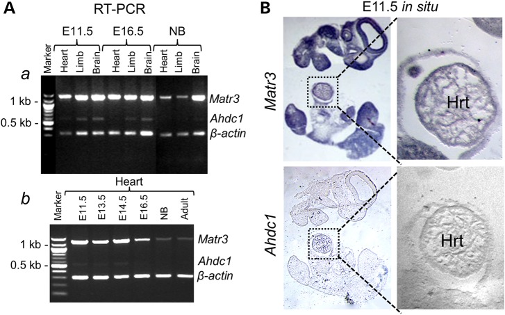
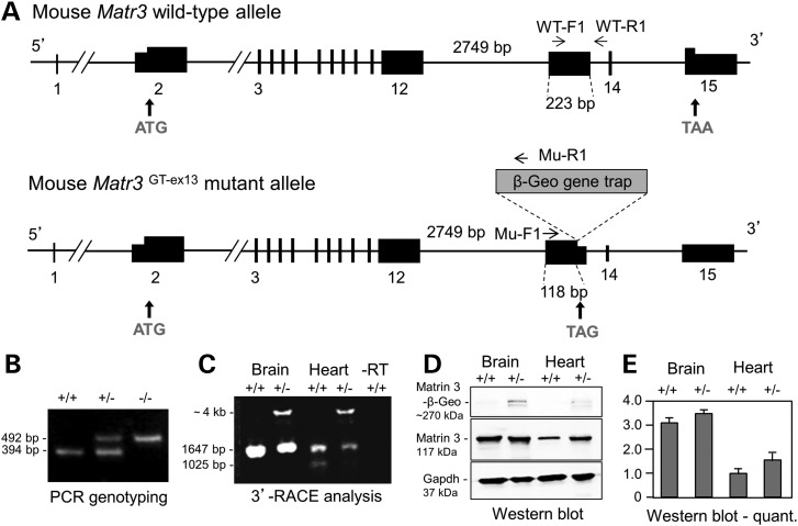
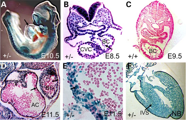
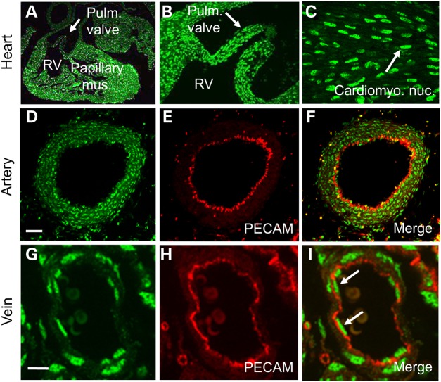
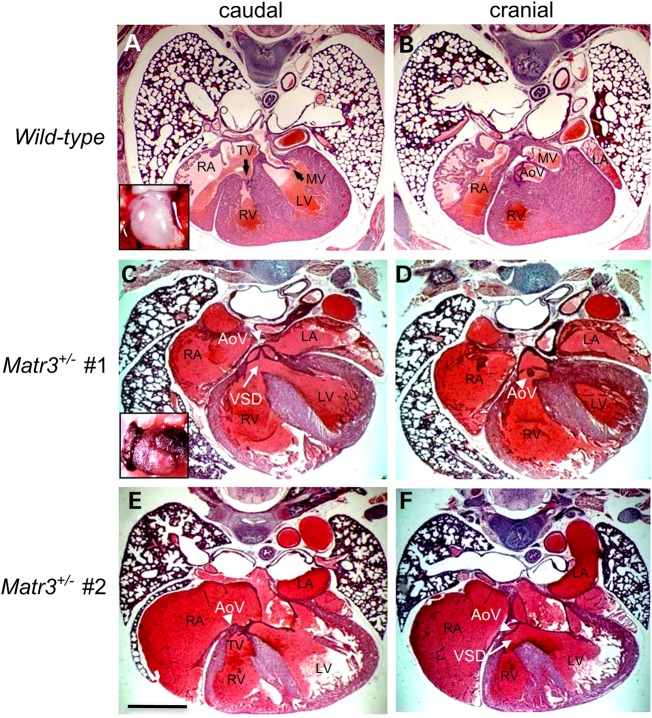
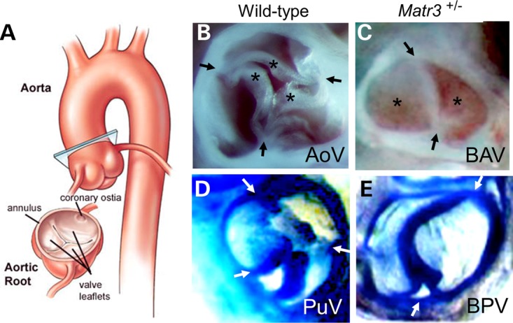
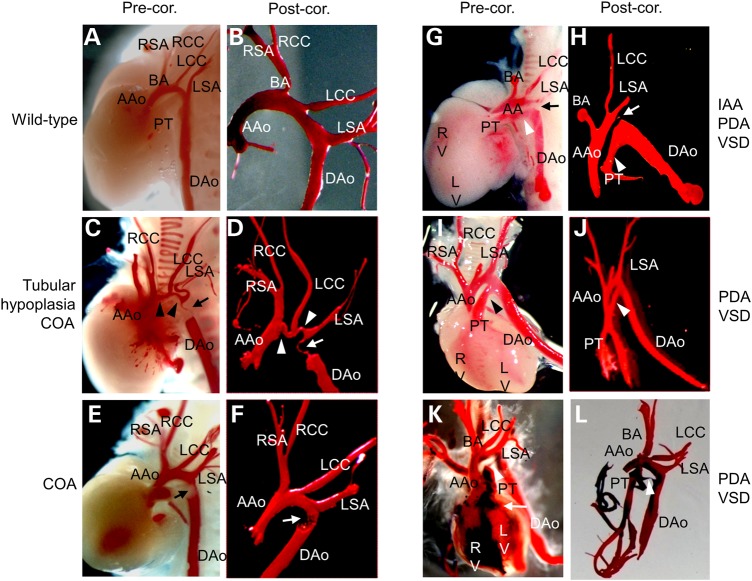
Cardiac left ventricular outflow tract (LVOT) defects represent a common but heterogeneous subset of congenital heart disease for which gene identification has been difficult. We describe a 46,XY,t(1;5)(p36.11;q31.2)dn translocation carrier with pervasive developmental delay who also exhibited LVOT defects, including bicuspid aortic valve (BAV), coarctation of the aorta (CoA) and patent ductus arteriosus (PDA). The 1p breakpoint disrupts the 5' UTR of AHDC1, which encodes AT-hook DNA-binding motif containing-1 protein, and AHDC1-truncating mutations have recently been described in a syndrome that includes developmental delay, but not congenital heart disease [Xia, F., Bainbridge, M.N., Tan, T.Y., Wangler, M.F., Scheuerle, A.E., Zackai, E.H., Harr, M.H., Sutton, V.R., Nalam, R.L., Zhu, W. et al. (2014) De Novo truncating mutations in AHDC1 in individuals with syndromic expressive language delay, hypotonia, and sleep apnea. Am. J. Hum. Genet., 94, 784-789]. On the other hand, the 5q translocation breakpoint disrupts the 3' UTR of MATR3, which encodes the nuclear matrix protein Matrin 3, and mouse Matr3 is strongly expressed in neural crest, developing heart and great vessels, whereas Ahdc1 is not. To further establish MATR3 3' UTR disruption as the cause of the proband's LVOT defects, we prepared a mouse Matr3(Gt-ex13) gene trap allele that disrupted the 3' portion of the gene. Matr3(Gt-ex13) homozygotes are early embryo lethal, but Matr3(Gt-ex13) heterozygotes exhibit incompletely penetrant BAV, CoA and PDA phenotypes similar to those in the human proband, as well as ventricular septal defect (VSD) and double-outlet right ventricle (DORV). Both the human MATR3 translocation breakpoint and the mouse Matr3(Gt-ex13) gene trap insertion disturb the polyadenylation of MATR3 transcripts and alter Matrin 3 protein expression, quantitatively or qualitatively. Thus, subtle perturbations in Matrin 3 expression appear to cause similar LVOT defects in human and mouse.


Figures

Figure 1.
Figure 1.
Clinical features, cardiac phenotype and translocation breakpoint structure in DGAP105. (A) Facial features at 4 years of age included mild hypertelorism, bilateral epicanthal folds, downslanting palpebral fissures, strabismus and a broad nose with a smooth philtrum and thin vermillion border. (B) Ideograms depicting 46,XY,t(1;5)(p36.11;q31.2)dn. Arrows mark locations of AHDC1 and MATR3 breakpoints. (C and D) Echocardiograms at age of 2 days. (C) Ductal view showing distal aortic arch, CoA just distal to the left subclavian artery (LSCA), accompanied by a prominent posterior unfolding (‘posterior shelf’) and PDA. (D) Aortic arch view showing ascending aorta (5.7-mm diameter), hypoplastic aortic arch (4.4-mm diameter) and CoA posterior shelf. (E) Summary of the 1p36.11 and 5q31.2 breakpoints in DGAP105. The 1p36.11 breakpoint disrupts AHDC1 intron 1, whereas the 5q31.2 breakpoint disrupts MATR3 exon 15 in the 3′ UTR. BACs used in FISH analyses are indicated.
Figure 2.
Figure 2.
Analysis of human MATR3 transcripts and protein expression in control and DGAP105 lymphoblasts. (A) Schematic of the MATR3 exon 13–15 region with the chromosomal translocation breakpoint in patient DGAP105 marked by dotted line. The proximal AAUAAA polyadenylation signal and the distal AAUAAA polyadenylation signal site are shown flanking the breakpoint in the 3′ UTR. TAA denotes the stop codon in exon 15. (B) MATR3 3′ RACE products in human control (Lane 1) and DGAP105 lymphoblast (Lanes 2, 3), and control human fetal heart tissue (Lanes 4, 5). RT ‘+’ or ‘−’ denote inclusion or omission of reverse transcriptase in the cDNA synthesis. The large product (1589 bp, arrow) uses the distal polyadenylation signal and predominates in control human lymphoblasts (Lane 1). In contrast, the short product (963 bp, arrow) predominates in DGAP105 lymphoblasts (Lane 2) and in control human fetal heart (Lane 4) and represents MATR3 transcripts that use the proximal polyadenylation signal. (C) Northern blot analysis of adult human tissues shows MATR3 transcripts of ∼3.5 and ∼2.9 kb. In heart and skeletal muscle, the 2.9-kb transcript predominates and likely corresponds to the 3′ RACE product using the proximal polyadenylation signal. In brain and other tissues, the 3.5-kb transcript predominates and corresponds to the 3′ RACE product using the distal polyadenylation signal. (D) Western blot analysis of protein isolated from DGAP105 and three control lymphoblast lines, showing up-regulation of Matrin 3 in DGAP105 compared with controls. Gapdh was used as loading control. (E) Quantification of Matrin 3 protein expression in D. Bars represent the mean fold expression of four independent experiments ± SEM, corrected for loading, and normalized to Control 2; *P < 0.05 between DGAP105 and mean of the three control lines via paired Student's t test.
Figure 3.
Figure 3.
Analysis of mouse Matr3 and Ahdc1 transcripts expression in developing heart. (A) RT–PCR analyses of Matr3 and Ahdc1. (a) Semi-quantitative RT–PCR analyses show strong Matr3 expression in developing mouse heart, limb and brain at E11.5, 16.5 stages, (b) with down-regulation at the newborn (NB) and adult stages. In contrast, Ahdc1 expression is only weakly detected in limb and brain at E11.5 and 16.5. (B) Section in situ hybridizations at E11.5 for mouse Matr3 and Ahdc1. Matr3 is expressed in CNS, pharyngeal arches, limb buds and in the developing heart (enlarged section), whereas Ahdc1 expression was undetectable in heart (enlarged section). Sense controls (not shown) showed no expression.
Figure 4.
Figure 4.
Analysis of the Matr3Gt-ex13 gene trap allele. (A) Structure of mouse Matr3 wild-type and Gt-ex13 gene trap mutant alleles. Wild-type mouse Matr3 encodes an 846-amino acid protein. Intron 12 (2749 bp) and exon 13 (223 bp) are shown. Matr3Gt-ex-13 gene trap allele inserts a β-Geo gene trap vector at position 118 bp in exon 13, which will generate Matr3-β-geo fusion transcripts. PCR genotyping primers depict WT-F1 (in exon 13) and WT-R1 (in intron 13) for the wild-type allele, and Mu-F1 (in exon 13) and Mu-R1 (in gene trap vector) for the mutant allele. Primers used in 3′ RACE are summarized on Materials and Methods. (B) E3.5 PCR genotyping shows 394-bp wild-type and 492-bp mutant alleles for wild-type (+/+), heterozygous (+/−) and homozygous (−/−) embryos. (C) Matr3GT-ex13 3′ RACE analysis of mouse E14.5 brain and heart tissues detects a novel Matr3-β-Geo fusion transcript (∼4 kb) in heterozygotes. The long Matr3 3′ RACE product (1647 bp), the only form detected in brain, is reduced in heterozygous brain. Both long and short Matr3 3′ RACE products (1647 and 1025 bp) are reduced in heterozygous heart. (D) Western blot analysis of Matrin 3 protein isolated from wild-type and heterozygous mouse E15.5 brain and heart tissues. Gapdh was used as loading control. (E) Quantification of Matrin 3 protein expression in D. Bars are fold ± SEM expression level from mean of three independent experiments, corrected for loading, and normalized to a value of 1.0 for wild-type heart. The small increase in expression in Matr3Gt-ex13/+ heart is not statistically significant.
Figure 5.
Figure 5.
X-Gal staining of Matr3Gt-ex13 heterozygotes. (A) X-Gal staining of primitive heart (arrow) in E10.5 Matr3 heterozygote, and in CNS (brain, spinal cord), pharyngeal arches and limb bud. (B) X-Gal staining in primitive heart of E8.5 Matr3 heterozygote. Arrow depicts dorsal mesocardium; BC, bulbus cordis; CVC, common ventricular chamber. (C) Negative control wild-type E9.5 embryo with eosin counterstain. (D) X-Gal staining in wall (arrow) of atrial chamber (AC), bulbus cordis (BC) and (E) myocardium and endocardium, and (F) interventricular septum (IVS) of newborn (NB) Matr3GT-ex13 heterozygote heart (arrows).
Figure 6.
Figure 6.
Matrin 3 protein cardiovascular expression in newborn mice. (A) Matrin 3 expression in wild-type newborn heart. (B) Pulmonary valve (PV) from (A) shows Matrin 3 in interstitial and endocardial cells. (C) Matrin 3 in cardiomyocyte nuclei (arrow). (D–F) Immunostaining in arterial vascular smooth muscle and endothelial cells for Matrin 3 (D), PECAM (E) and merged (F). Matrin 3 is expressed in both arterial smooth muscle cell nuclei (green) external to PECAM-1 endothelial cell membrane staining (red) and internal to the PECAM-1 domain, in endothelial cell nuclei. This is better seen in (G–I) with Matrin 3 and PECAM in small venules, which are largely devoid of vascular smooth muscle. Arrows in (I) denote Matrin 3 in endothelial cell nuclei. Scale bar: 40 μm (D–F); 5 μm (G–I).
Figure 7.
Figure 7.
Subaortic VSD and DORV phenotypes in Matr3Gt-ex13 heterozygotes. Transverse serial sections through the hearts of E18.5 wild type (A and B), and two different representative Matr3Gt-ex13 heterozygotes (embryo 1 in C and D and embryo 2 in E and F), each sectioned at a cranial and caudal level, illustrating the DORV with subaortic VSD phenotype in Matr3Gt-ex13 heterozygotes (see Table 2). (A and B) Wild-type sections show left and right ventricles separated by the interventricular septum, the two atrioventricular (tricuspid, mitral) valves and the aortic valve (pulmonic valve not seen in this view). Heterozygous sections do not closely resemble wild-type sections in overall cardiac configuration because, in addition to specific cardiac anomalies, affected newborn Matr3GT-ex13 heterozygote hearts are frequently maloriented and exhibit an abnormal ‘boot shape’, with the cardiac apex pointing horizontally to the animal's left (see insets, A and C). (C) Subaortic VSD is directly inferior to and aligned with the aortic valve. (D) The aortic valve significantly overrides the right ventricle, which together with the normal communication of right ventricle to pulmonary artery (data not shown), establishes DORV. (E) In this specimen, an unusually close continuity exists between the tricuspid and aortic valves. (F) Subaortic VSD and DORV are shown. AoV, aortic valve; LA and LV, left atrium and ventricle; MV, mitral valve; RA and RV, right atrium and ventricle; TV, tricuspid valve; VSD, ventricular septal defect. Scale bar: 500 μm.
Figure 8.
Figure 8.
Semilunar heart valve defects in Matr 3Gt-ex-13 heterozygotes. (A) Diagram of ascending aorta, aortic arch and descending aorta, showing plane of section for aortic valve analysis. Note left and right coronary ostia (openings) below the fibrous annulus (ring) that demarcates the valve [Cleveland Clinic Foundation (CCF), with permission]. (B) Wild-type newborn aortic valve (AoV) showing tri-leaflet (*) morphology in open configuration. Commissural attachments to the annulus are marked (arrows). (C) Matr3GT-ex13 heterozygote newborn bicuspid AoV (BAV) showing two leaflets in closed configuration. (D) Wild-type newborn pulmonic valve (PuV) showing tri-leaflet morphology in open configuration. (E) Matr3GT-ex13 heterozygote newborn bicuspid PuV (BPV) showing two leaflets in closed configuration.
Figure 9.
Figure 9.
Aortic arch abnormalities in Matr3Gt-ex13 heterozygotes. (A and B) Newborn wild-type aortic arch vasculature, showing pre- (A) and post-corrosion (B) cast analysis. (C–L) Matr3 heterozygous newborns with various outflow tract defects. (C and D) Tubular hypoplasia and CoA. The deformed aortic arch is uniformly narrowed (segment between arrowheads), and a CoA (arrow) lies distal to the LSA near a closed DAo. (E and F) CoA (arrow) just distal to the LSA and at the level of the closed DAo also called a ‘juxaductal CoA’. (G and H) Interrupted aortic arch just distal to the LSA, with a strand of tissue joining the two segments (‘atretic aortic arch’; arrow). A VSD with left to right shunting is also present, as evident by red polymer in both ventricles. A large PDA (arrowhead) is the sole source of blood to the lower half of the body. (I and J) A wide PDA (arrowhead) and VSD are present. Following LV injection, both ventricles and the PT contain red polymer; the PT is connected to the PDA that joins the DAo. (K and L) Dual-color corrosion casting shows admixture of red (injected into LV) and blue polymers (injected into RV) in both ventricles, confirming the presence of a VSD (arrow, K). Both polymers are also present in the pulmonary trunk and aorta. A small PDA is present (arrowheads, K and L). AAo, ascending aorta; BA, brachiocephalic artery; DAo, descending aorta; IAA, interrupted aortic arch; LV, left ventricle; PT, pulmonary trunk; RCC/LCC, right/left common carotid arteries; RSA/LSA, right/left subclavian arteries; RV, right ventricle; VSD, ventricular septal defect.

References

    1. Andersen T.A., Troelsen Kde L., Larsen L.A. (2014) Of mice and men: molecular genetics of congenital heart disease. Cell. Mol. Life Sci., 71, 1327–1352. - PMC - PubMed
    1. Lalani S.R., Belmont J.W. (2014) Genetic basis of congenital cardiovascular malformations. Eur. J. Med. Genet., 57, 402–413. - PMC - PubMed
    1. Jenkins K.J., Correa A., Feinstein J.A., Botto L., Britt A.E., Daniels S.R., Elixson M., Warnes C.A., Webb C.L. (2007) Noninherited risk factors and congenital cardiovascular defects: current knowledge: a scientific statement from the American Heart Association Council on Cardiovascular Disease in the Young: endorsed by the American Academy of Pediatrics. Circulation, 115, 2995–3014. - PubMed
    1. Pierpont M.E., Basson C.T., Benson D.W., Jr, Gelb B.D., Giglia T.M., Goldmuntz E., McGee G., Sable C.A., Srivastava D., Webb C.L. (2007) Genetic basis for congenital heart defects: current knowledge: a scientific statement from the American Heart Association Congenital Cardiac Defects Committee, Council on Cardiovascular Disease in the Young: endorsed by the American Academy of Pediatrics. Circulation, 115, 3015–3028. - PubMed
    1. Siu S.C., Silversides C.K. (2010) Bicuspid aortic valve disease. J. Am. Coll. Cardiol., 55, 2789–2800. - PubMed

Publication types

MeSH terms