Review
. 2015 Jan 7;21(1):60-83.
doi: 10.3748/wjg.v21.i1.60.

Autoimmune hepatitis, one disease with many faces: etiopathogenetic, clinico-laboratory and histological characteristics

Affiliations
Review

Autoimmune hepatitis, one disease with many faces: etiopathogenetic, clinico-laboratory and histological characteristics

Nikolaos K Gatselis et al. World J Gastroenterol. .

Abstract

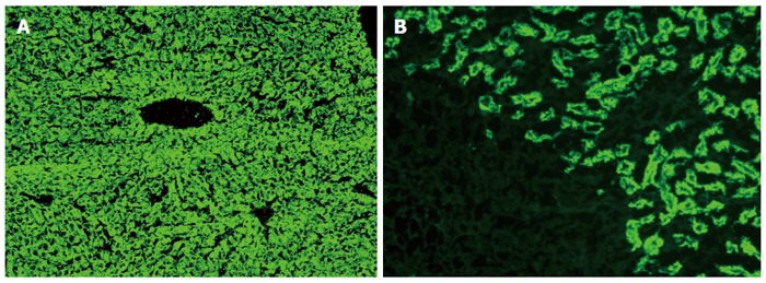
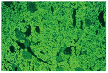
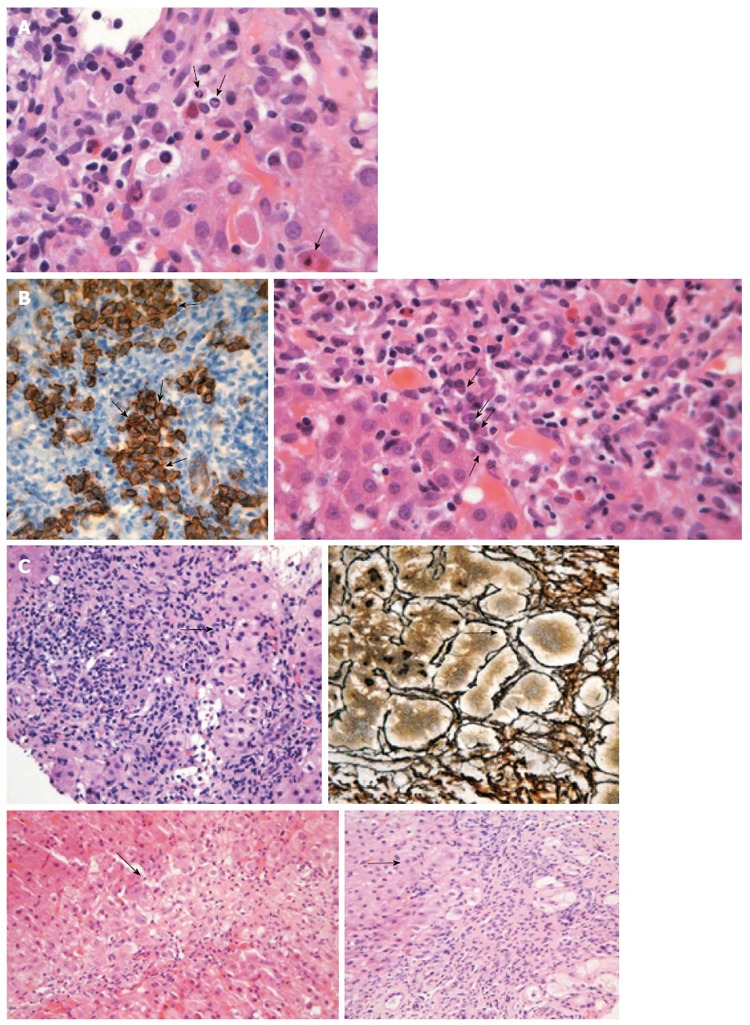
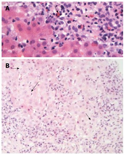
Autoimmune hepatitis (AIH) is an unresolving progressive liver disease of unknown etiology characterized by hypergammaglobulinemia, autoantibodies detection and interface hepatitis. Due to the absence of specific diagnostic markers and the large heterogeneity of its clinical, laboratory and histological features, AIH diagnosis may be potentially difficult. Therefore, in this in-depth review we summarize the substantial progress on etiopathogenesis, clinical, serological and histological phenotypes of AIH. AIH has a global distribution affecting any age, both sexes and all ethnic groups. Clinical manifestations vary from asymptomatic to severe or rarely fulminant hepatitis. Hypergammaglobulinemia with selective elevation of IgG is found in most cases. Autoimmune attack is perpetuated, possibly via molecular mimicry, and favored by the impaired control of T-regulatory cells. Histology (interface hepatitis, emperipolesis and hepatic rosette formation) and autoantibodies detection although not pathognomonic, are still the hallmark for a timely diagnosis. AIH remains a major diagnostic challenge. AIH should be considered in every case in the absence of viral, metabolic, genetic and toxic etiology of chronic or acute hepatitis. Laboratory personnel, hepato-pathologists and clinicians need to become more familiar with disease expressions and the interpretation of liver histology and autoimmune serology to derive maximum benefit for the patient.

Keywords: Autoimmune hepatitis; Liver autoimmunity; Liver-related autoantibodies; Non-organ specific autoantibodies; Overlap syndromes.


Figures

Figure 1
Figure 1
Typical diffuse staining of the cytoplasm of the entire liver lobule on rat liver sections in a case of autoimmune hepatitis type 2 positive for antibodies against liver-kidney microsomes type 1 (A); Antibodies against liver-kidney microsomes type 1 react to the proximal tubules of the rat kidney and the absence of reactivity against the distal tubules of the rat kidney (see also Figure 2) and parietal cells of the rat stomach distinguishes anti-LKM1 autoantibodies from antimitochondrial antibodies (B). Original magnification × 40.
Figure 2
Figure 2
Contrary to antibodies against liver-kidney microsomes type 1 which react to the proximal tubules, antimitochondrial antibodies react to the proximal and also the distal tubules of the rat kidney (original magnification × 40). In these cases there is also reactivity to the parietal cells of the rat stomach.
Figure 3
Figure 3
Liver histology in autoimmune hepatitis. A: Interface hepatitis with genuine apoptotic bodies (arrows) in a case with autoimmune hepatitis-type 1 (hematoxylin and eosin staining). See also the typical rim of condensed chromatin at the nuclear membrane and ordinary acidophilic bodies, at various stages of development; B: Intense portal plasmacytosis in autoimmune hepatitis (arrows in the right panel; hematoxylin and eosin staining). The plasma cells are in clusters, better seen after immunostaining with CD138 (arrows in dark brown staining of the left panel). The intensity of plasmacytosis can be useful in discriminating autoimmune hepatitis from viral hepatitis whereas there is also possible prognostic information as portal plasma cell infiltrates while on immunosuppression are associated with relapse upon drug cessation; C: Various examples of rosette formation (hematoxylin and eosin staining; periportal, periseptal in a previous collapse and with reticulin staining seen in the upper right panel; see arrows).
Figure 4
Figure 4
Liver histology in autoimmune hepatitis (continued). A: In some cases of autoimmune hepatitis, “numerous” eosinophils or eosinophils as a minor component of the inflammatory infiltrate can be seen (arrows; hematoxylin and eosin staining). This is an interesting and useful observation, since this finding is evaluated falsely out of the histopathological context of autoimmune hepatitis, and therefore, could make the differential diagnosis from drug-induced hepatitis more problematic; B: Interface hepatitis with hydropic change of involved hepatocytes (arrows; hematoxylin and eosin staining). Note the presence of adjacent parenchymal collapse, alternatively multiacinar necrosis, which has been considered as one of the most distinguishing morphological features of autoimmune hepatitis since its presence in the appropriate clinicopathological context is supportive of the diagnosis.

References

    1. Zachou K, Muratori P, Koukoulis GK, Granito A, Gatselis N, Fabbri A, Dalekos GN, Muratori L. Review article: autoimmune hepatitis -- current management and challenges. Aliment Pharmacol Ther. 2013;38:887–913. - PubMed
    1. Dalekos GN, Zachou K, Liaskos C, Gatselis N. Autoantibodies and defined target autoantigens in autoimmune hepatitis: an overview. Eur J Intern Med. 2002;13:293–303. - PubMed
    1. Czaja AJ. Autoantibodies in autoimmune liver disease. Adv Clin Chem. 2005;40:127–164. - PubMed
    1. Krawitt EL. Can you recognize autoimmune hepatitis? Postgrad Med. 1998;104:145–149, 152. - PubMed
    1. Johnson PJ, McFarlane IG. Meeting report: International Autoimmune Hepatitis Group. Hepatology. 1993;18:998–1005. - PubMed

LinkOut - more resources