. 2014:2014:657876.
doi: 10.1155/2014/657876. Epub 2014 Dec 9.

Cutaneous angiosarcoma of the foot: a case report and review of the literature

Affiliations

Cutaneous angiosarcoma of the foot: a case report and review of the literature

Sharang Tenjarla et al. Case Rep Oncol Med. 2014.

Abstract

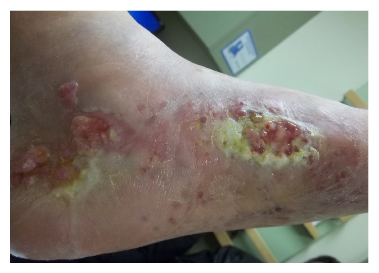
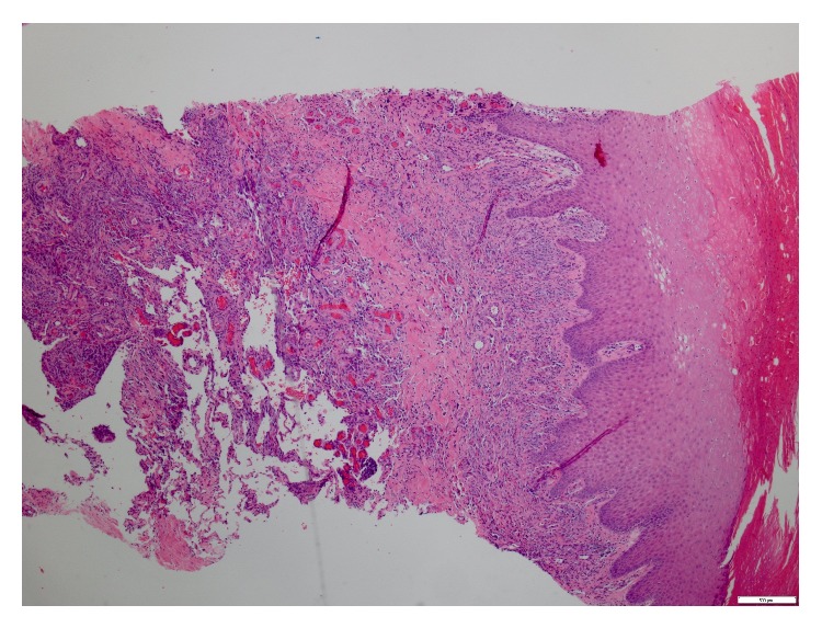
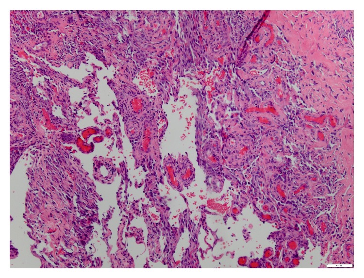
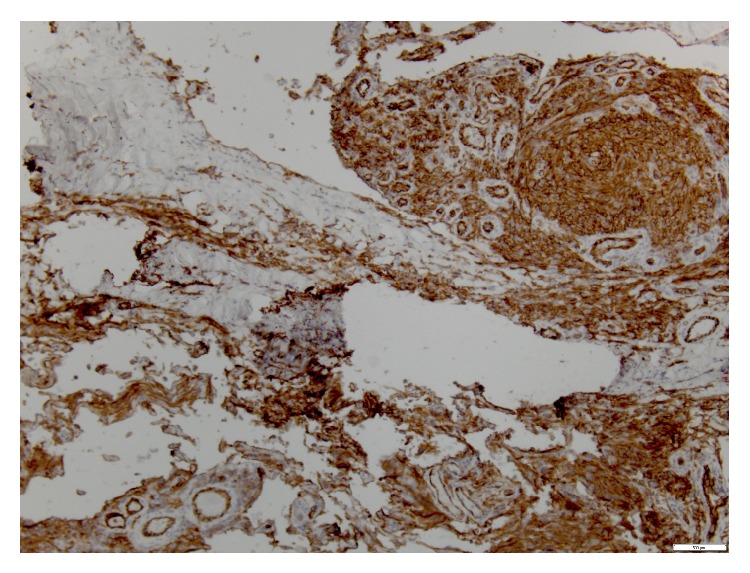
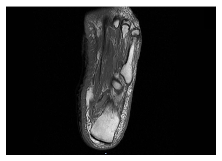
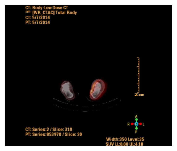
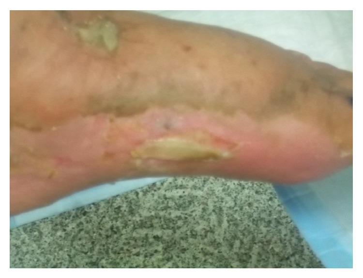
Primary Angiosarcoma of the skin of the foot is very rare. Angiosarcoma is typically treated with resection and wide-field postoperative radiation therapy. Chemotherapy and radiation therapy have also been used. Regardless of the treatment, the risk of local and distant relapse remains high for this disease. We present a case of an elderly patient who developed cutaneous angiosarcoma of the foot. It posed as a diagnostic dilemma at presentation. Chronic lymphedema was a possible predisposing factor. Given his age, preexisting renal dysfunction, refusal of surgery, and preference not to receive chemotherapy, the patient was ultimately treated with definitive radiotherapy. We present this case because of its rare site, unique presentation and delay in diagnosis of the condition, and attainment of an excellent response to radiation at the time of follow-up. We also review the current literature on this topic.


Figures

Figure 1
Figure 1
Pretreatment: 5 × 5 cm ulcer on the medial left foot emanating a serosanginous discharge and similar ulcer present posteriorly along with blistering satellite lesions on the plantar surface of the foot.
Figure 2
Figure 2
Low power view showing extensive spindle cell proliferation involving dermis, subcutis, and deeper fibroadipose tissue.
Figure 3
Figure 3
High power view showing multiple vascular sinuses lined by tufts of neoplastic endothelial cells.
Figure 4
Figure 4
Positivity for vascular marker CD-31.
Figure 5
Figure 5
MRI of the left foot with contrast showing diffuse soft tissue T1 hypointense signal within both medial and lateral subcutaneous tissues, which is more prominent in the fat anterior to the Achilles tendon.
Figure 6
Figure 6
FDG-PET and CT scan showing heterogeneous uptake in the medial and lateral foot with a more focal uptake in the medial foot, anterior to the Achilles tendon.
Figure 7
Figure 7
Two months postradiation: marked diminishment of cutaneous ulceration with decline in discharge and drying of satellite nodules.

References

    1. Mark R. J., Poen J. C., Tran L. M., Fu Y. S., Juillard G. F. Angiosarcoma: a report of 67 patients and a review of the literature. Cancer. 1996;77(11):2400–2406. - PubMed
    1. Weiss S. W., Goldblum J. R., Folpe A. L. Enzinger and Weiss's Soft Tissue Tumors. St. Louis, Mo, USA: Mosby; 2007.
    1. Penel N., Lansiaux A., Adenis A. Angiosarcomas and taxanes. Current Treatment Options in Oncology. 2007;8(6):428–434. doi: 10.1007/s11864-007-0042-0. - DOI - PubMed
    1. Trinh N. Q., Rashed I., Hutchens K. A., Go A., Meilan E., Tung R. Unusual clinical presentation of cutaneous angiosarcoma masquerading as eczema: a case report and review of the literature. Case Reports in Dermatological Medicine. 2013;2013 doi: 10.1155/2013/906426.906426 - DOI - PMC - PubMed
    1. Holden C. A., Spittle M. F., Jones E. W. Angiosarcoma of the face and scalp, prognosis and treatment. Cancer. 1987;59(5):1046–1057. doi: 10.1002/1097-0142(19870301)59:5<1046::AID-CNCR2820590533>3.0.CO;2-6. - DOI - PubMed

LinkOut - more resources