. 2014:2014:523924.
doi: 10.1155/2014/523924.

Mechanisms of Myofascial Pain

Affiliations

Mechanisms of Myofascial Pain

M Saleet Jafri. Int Sch Res Notices. 2014.

Abstract

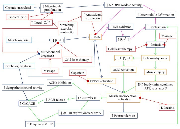
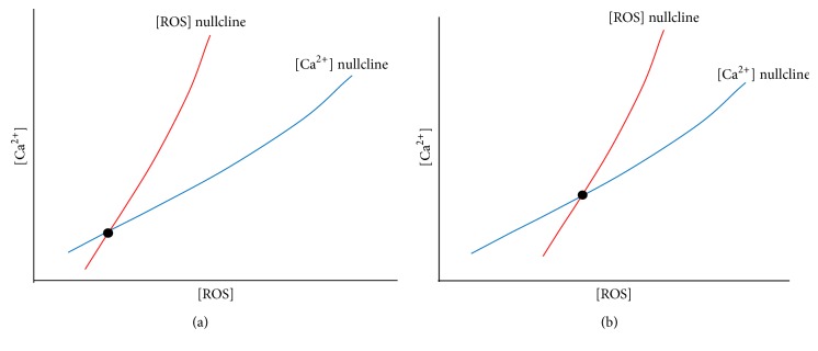
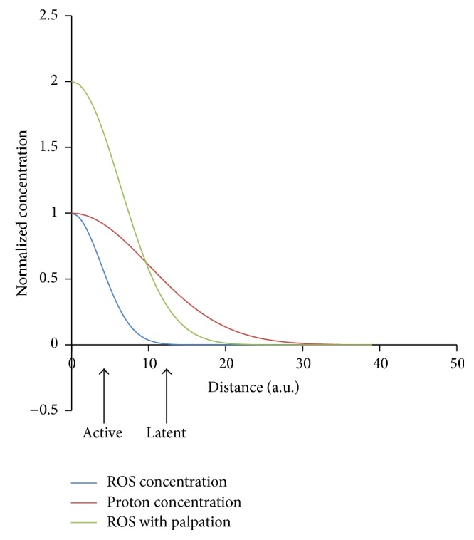
Myofascial pain syndrome is an important health problem. It affects a majority of the general population, impairs mobility, causes pain, and reduces the overall sense of well-being. Underlying this syndrome is the existence of painful taut bands of muscle that contain discrete, hypersensitive foci called myofascial trigger points. In spite of the significant impact on public health, a clear mechanistic understanding of the disorder does not exist. This is likely due to the complex nature of the disorder which involves the integration of cellular signaling, excitation-contraction coupling, neuromuscular inputs, local circulation, and energy metabolism. The difficulties are further exacerbated by the lack of an animal model for myofascial pain to test mechanistic hypothesis. In this review, current theories for myofascial pain are presented and their relative strengths and weaknesses are discussed. Based on new findings linking mechanoactivation of reactive oxygen species signaling to destabilized calcium signaling, we put forth a novel mechanistic hypothesis for the initiation and maintenance of myofascial trigger points. It is hoped that this lays a new foundation for understanding myofascial pain syndrome and how current therapies work, and gives key insights that will lead to the improvement of therapies for its treatment.


Figures

Figure 1
Figure 1
Hypothesized signaling pathways of myofascial trigger points and therapies. A schematic diagram of the mechanism of myofascial trigger points and treatment options. The initiating mechanisms of myofascial trigger points are shown in blue with the downstream events in dark blue. The treatment for myofascial pain is shown in red with the pathways of their action shown in dark red. The arrows indicate how one feature causes another. The square at the ends of lines indicates inhibition of the end feature by the preceding feature. The myofascial trigger point is initiated by a combination of chronic load on the muscle which caused microtubule proliferation which increases ROS production and a decreased ability to remove ROS. The increase of ROS increases ryanodine receptors open probability, hence increasing calcium which results in contraction and deformation of the microtubule network resulting in more ROS production. This is the key positive feedback loop. Psychological stress can contribute to this as it reduces mitochondrial content and increase ROS production in cells. The contraction restricts blood flow resulting in local ischemia/hypoxia that results in muscle damage and the inflammatory response. Pain is caused by the activation of nociceptors by a decreased pH (ASIC channels), increased ROS (TRPV1 channels), and substance P. When depolarized, nociceptive neurons release CGRP which increases the amount of and response to acetylcholine in the neuromuscular junction, which can cause additional contraction. This provided a second positive feedback loop. Treatments such as lidocaine and capsaicin block nociception. Other treatments such as massage and cold laser therapy might increase circulation or reduce oxidative stress. Furthermore, treatments that induce stretching/contraction such as needling, electrical stimulation, stretching, and exercise increase calcium local to high enough concentrations (>10 μM) transiently that induce microtubule depolymerization. Finally, the application of topical or injected thiocolchicine has been shown to reduce myofascial pain. Abbreviations: GSH (glutathione), ROS (reactive oxygen species), NADPH (nicotinamide adenine dinucleotide phosphate), RyR (ryanodine receptors), [Ca2+] (calcium concentration), [H+] (proton concentration), ATP (adenosine triphosphate), TRPV1 (transient receptor potential cation channel subfamily V member 1 or capsaicin receptor), ASIC (acid-sensing ion channel), CGRP (calcitonin gene related peptide), ACH (acetylcholine), ACHe (acetylcholinesterase), and ACHR (acetylcholine receptor).
Figure 2
Figure 2
Hypothesized mechanism yielding active and latent trigger points. At the site of the myofascial trigger point, there is restriction of blood flow increasing the proton concentration ([H+]) locally. There is also an increase of local [ROS]. The plot shows the normalized concentrations of these substances as a function of the distance from the trigger point center. As the distance from the trigger point center increases, the levels of [ROS] (blue) decrease more gradually than the levels of [H+] (red). If the nociceptors are located close to the center of the trigger point, they are active as the levels of [ROS] and [H+] protons are high enough to activate the ASIC and TRPV1 channels. If these neurons/receptors are farther away the, trigger point would be latent. Upon palpation the [ROS] increases due to mechanical deformation of the microtubule network and activation of NADPH oxidase and the [ROS] increases (green) so that the nociceptors see sufficient ROS to be activated.
Figure 3
Figure 3
Hypothesis of how changes to the affected region cause a sustained perturbation yielding a trigged point. Conceptual phase plane diagrams for intracellular [Ca2+] and [ROS]. (a) The [ROS] nullcline (red) intersects the [Ca2+] nullcline (blue) at the steady state value for resting [Ca2+] and [ROS] (black dot). (b) With the changes that occur with microtubule proliferation and reduction in the ability of the myocyte to remove ROS, the [ROS] nullcline shifts to the right higher [ROS] for a given level of [Ca2+] resulting in a higher steady-state [ROS] and [Ca2+].

References

    1. Simons D. G. Clinical and etiological update of myofascial pain from trigger points. Journal of Musculoskeletal Pain. 1996;4(1-2):93–121. doi: 10.1300/J094v04n01_07. - DOI
    1. Fleckenstein J., Zaps D., Rüger L. J., et al. Discrepancy between prevalence and perceived effectiveness of treatment methods in myofascial pain syndrome: results of a cross-sectional, nationwide survey. BMC Musculoskeletal Disorders. 2010;11, article 32 doi: 10.1186/1471-2474-11-32. - DOI - PMC - PubMed
    1. Gerber L. H., Sikdar S., Armstrong K., et al. A systematic comparison between subjects with no pain and pain associated with active myofascial trigger points. PM & R: The Journal of Injury, Function, and Rehabilitation. 2013;5(11):931–938. doi: 10.1016/j.pmrj.2013.06.006. - DOI - PMC - PubMed
    1. Simons D. G. Review of enigmatic MTrPs as a common cause of enigmatic musculoskeletal pain and dysfunction. Journal of Electromyography and Kinesiology. 2004;14(1):95–107. doi: 10.1016/j.jelekin.2003.09.018. - DOI - PubMed
    1. Simons D. G ., Travell J. G., Simons L. S., Cummings B. D. Travell & Simons' Myofascial Pain and Dysfunction: The Trigger Point Manual. Lippincott Williams & Wilkins; 1998.

LinkOut - more resources