Review
. 2015 Feb 17;48(2):457-65.
doi: 10.1021/ar500340a. Epub 2015 Jan 9.

Enzyme dynamics from NMR spectroscopy

Affiliations
Review

Enzyme dynamics from NMR spectroscopy

Arthur G Palmer 3rd. Acc Chem Res. .

Abstract

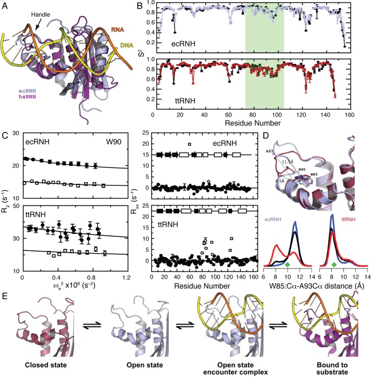
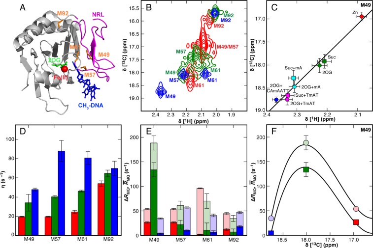
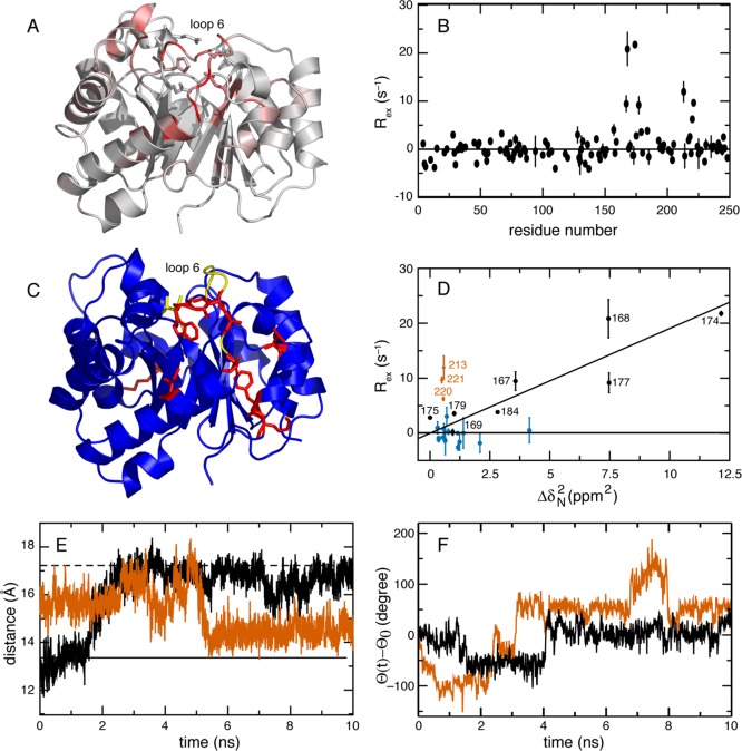
CONSPECTUS: Biological activities of enzymes, including regulation or coordination of mechanistic stages preceding or following the chemical step, may depend upon kinetic or equilibrium changes in protein conformations. Exchange of more open or flexible conformational states with more closed or constrained states can influence inhibition, allosteric regulation, substrate recognition, formation of the Michaelis complex, side reactions, and product release. NMR spectroscopy has long been applied to the study of conformational dynamic processes in enzymes because these phenomena can be characterized over multiple time scales with atomic site resolution. Laboratory-frame spin-relaxation measurements, sensitive to reorientational motions on picosecond-nanosecond time scales, and rotating-frame relaxation-dispersion measurements, sensitive to chemical exchange processes on microsecond-millisecond time scales, provide information on both conformational distributions and kinetics. This Account reviews NMR spin relaxation studies of the enzymes ribonuclease HI from mesophilic (Escherichia coli) and thermophilic (Thermus thermophilus) bacteria, E. coli AlkB, and Saccharomyces cerevisiae triosephosphate isomerase to illustrate the contributions of conformational flexibility and dynamics to diverse steps in enzyme mechanism. Spin relaxation measurements and molecular dynamics (MD) simulations of the bacterial ribonuclease H enzymes show that the handle region, one of three loop regions that interact with substrates, interconverts between two conformations. Comparison of these conformations with the structure of the complex between Homo sapiens ribonuclease H and a DNA:RNA substrate suggests that the more closed state is inhibitory to binding. The large population of the closed conformation in T. thermophilus ribonuclease H contributes to the increased Michaelis constant compared with the E. coli enzyme. NMR spin relaxation and fluorescence spectroscopy have characterized a conformational transition in AlkB between an open state, in which the side chains of methionine residues in the active site are disordered, and a closed state, in which these residues are ordered. The open state is highly populated in the AlkB/Zn(II) complex, and the closed state is highly populated in the AlkB/Zn(II)/2OG/substrate complex, in which 2OG is the 2-oxoglutarate cosubstrate and the substrate is a methylated DNA oligonucleotide. The equilibrium is shifted to approximately equal populations of the two conformations in the AlkB/Zn(II)/2OG complex. The conformational shift induced by 2OG ensures that 2OG binds to AlkB/Zn(II) prior to the substrate. In addition, the opening rate of the closed conformation limits premature release of substrate, preventing generation of toxic side products by reaction with water. Closure of active site loop 6 in triosephosphate isomerase is critical for forming the Michaelis complex, but reopening of the loop after the reaction is (partially) rate limiting. NMR spin relaxation and MD simulations of triosephosphate isomerase in complex with glycerol 3-phosphate demonstrate that closure of loop 6 is a highly correlated rigid-body motion. The MD simulations also indicate that motions of Gly173 in the most flexible region of loop 6 contribute to opening of the active site loop for product release. Considered together, these three enzyme systems illustrate the power of NMR spin relaxation investigations in providing global insights into the role of conformational dynamic processes in the mechanisms of enzymes from initial activation to final product release.


Figures

Figure 1
Figure 1
Autoinhibition of RNase H., (A) Superposition of the ecRNH structure (light blue, PDB ID 2RN2) with the substrate-bound complex of the hsRNH protein (purple; PDB ID 2QK9), illustrating the position of the handle region interacting with the (yellow) DNA strand of the DNA:RNA hybrid substrate. (B) Backbone amide 15N S2 (ecRNH residue numbering is used throughout this figure). (top) Experimental (black) and predicted (blue) S2 for ecRNH. (bottom) Experimental (black) and predicted (red) S2 for ttRNH. Helices B and C and the handle region are highlighted in green. Experimental values are rescaled by linear regression to the simulated values for visualization. (C) R relaxation dispersion for backbone amide 15N nuclei. Trp90 R2e) is shown for (top left) ecRNH and (bottom left) ttRNH at 300 K. Closed and open symbols represent data collected at 11.7 and 18.8 T, respectively. Backbone amide 15N Rex at 300 K, 14.1 T for (top right) ecRNH and (bottom right) ttRNH. Values of Rex ≥ 2.5 s–1 are indicated by open circles. (D) (top) Representative conformations from an ecRNH MD trajectory of the (blue) open and (brown) closed states, illustrating the Cartesian W85-Cα–A93-Cα distance metric used to distinguish open (∼11.5 Å) and closed conformations (∼8.5 Å). The location of Trp90 also is shown. (bottom) Temperature dependence of ecRNH and ttRNH conformational distributions, illustrating the relative populations of the closed and open handle-region states at (blue) 273 K, (black) 300 K, and (red) 340 K. Values of the distance metric from ecRNH and ttRNH (PDB ID 1RIL) crystal structures are shown as green diamonds. (E) Kinetic scheme for the interaction of substrate with the handle region of RNase H, in which the closed and open states are incompetent and competent for binding, respectively. Panel C is from ref (38); other panels are from ref (39).
Figure 2
Figure 2
Conformation and dynamics of AlkB characterized by 1H–13C NMR at 283 K. (A) X-ray crystal structure of AlkB (PDB ID 2FD8). The Fe(II)/2OG core and NRL are colored gray and magenta, respectively; substrates and cosubstrates (red) Fe(II), (green) 2OG, and (blue) 5′-TmAT-3′ (mA = 1-methyl A) are depicted as spheres and sticks; methionine residues are shown as orange sticks. (B) Methyl 1H–13C spectra for 13Cε-Met AlkB successively titrated with (red) 2.0 × Zn(II), (green) 10.0 × 2OG cosubstrate, and (blue) 1.5 × 5′-CAmAAT-3′ substrate. (C) Linear correlations of the M49 chemical shifts from panel A and from additional spectra of the enzyme saturated with the alternative substrates 1-methyladenosine triphosphate (mA, cyan) or 5′-TmAT-3′ (TmAT, magenta). (D) The methyl 1H–1H cross-correlated relaxation rate η. An approximate value τc = 21.9 ± 0.5 ns yields a maximum value of η = 79 ± 2 s–1 for the closed AlkB/Zn(II)/2OG/5′-CAmCAT-3′ complex. This estimate suggests that M57 and M61 are highly immobilized in the closed complex. (E) (translucent) MQ = (RDQ + RZQ)/2 and (solid) ΔRMQ = (RDQRZQ)/2 for 13Cε-Met resonances in AlkB, colored as in panel B. RZQ and RDQ represent the zero- and double-quantum relaxation rate constants, respectively. (F) Two-state chemical exchange model (solid lines) for (circles) MQ or (squares) ΔMQ. This research was originally published in ref (44). Copyright 2014 American Society for Biochemistry and Molecular Biology.
Figure 3
Figure 3
Loop 6 dynamics in triosephosphate isomerase (TIM). (A) 15N (ΔδN) and 1H (ΔδH) chemical shift changes in TIM upon the binding of G3P. The values of (10ΔδH2 + ΔδN2)1/2 are color coded onto the structure of the TIM monomer from white (0 ppm) to red (7.1 ppm). Active site residues Asn10, Lys12, His95, Ser96, Glu97, and Glu165 are shown in stick representation. (B) Rex for G3P-bound TIM at 298 K and 18.8 T. (C) Cartoon representation of the apo structure of TIM; (red) residues with Rex > 0, and (yellow) residues located in loop 6 with Rex = 0. (D) Rex versus ΔδN2 at 298 K. A linear correlation is observed for loop 6 residues colored in black (correlation coefficient = 0.9). Rex for (vermillion) residues 213, 220, and 221 is not correlated with loop 6 closing. (blue) Residues that are not part of loop 6 with ΔδN2 > 0 but Rex ≈ 0. (E) Distance between the Cα atoms of Gly171 and Y208 monitors loop 6 opening and closing in MD simulations starting from (black) closed (PDB 7TIM) and (vermillion) open (PDB 1YPI) conformations performed at 300 K in the absence of bound ligands. Horizontal lines show the distances in the crystal structures of closed () and open (---) states. (F) Cα pseudodihedral angle, Θ, for Gly 173 for trajectories that started from the (black) closed and (vermillion) open states; Θ0 is the value of Θ in the corresponding X-ray structure. Adapted with permission from ref (52). Copyright 2006 American Chemical Society.

References

    1. Palmer A. G.NMR spectroscopy: NMR relaxation methods. In Biophysical Techniques for Structural Characterization of Macromolecules; Dyson H. J., Ed.; Academic Press: Oxford, 2012; Vol. 1; pp 216–244.
    1. Palmer A. G. Chemical exchange in biomacromolecules: Past, present, and future. J. Magn. Reson. 2014, 241, 3–17. - PMC - PubMed
    1. Palmer A. G.; Massi F. Characterization of the dynamics of biomacromolecules using rotating-frame spin relaxation NMR spectroscopy. Chem. Rev. 2006, 106, 1700–1719. - PubMed
    1. Clore G. M. Seeing the invisible by paramagnetic and diamagnetic NMR. Biochem. Soc. Trans. 2013, 41, 1343–1354. - PMC - PubMed
    1. Chen K.; Tjandra N. The use of residual dipolar coupling in studying proteins by NMR. Top. Curr. Chem. 2012, 326, 47–67. - PMC - PubMed

Publication types

LinkOut - more resources