. 2015 Jan 8;96(1):104-20.
doi: 10.1016/j.ajhg.2014.12.004.

Genome-wide comparative analysis of atopic dermatitis and psoriasis gives insight into opposing genetic mechanisms

Affiliations

Genome-wide comparative analysis of atopic dermatitis and psoriasis gives insight into opposing genetic mechanisms

Hansjörg Baurecht et al. Am J Hum Genet. .

Erratum in

  • Am J Hum Genet. 2015 Dec 3;97(6):933

Abstract

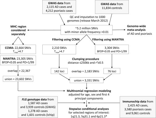
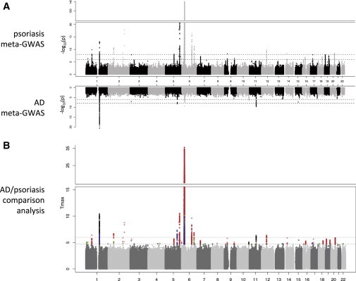
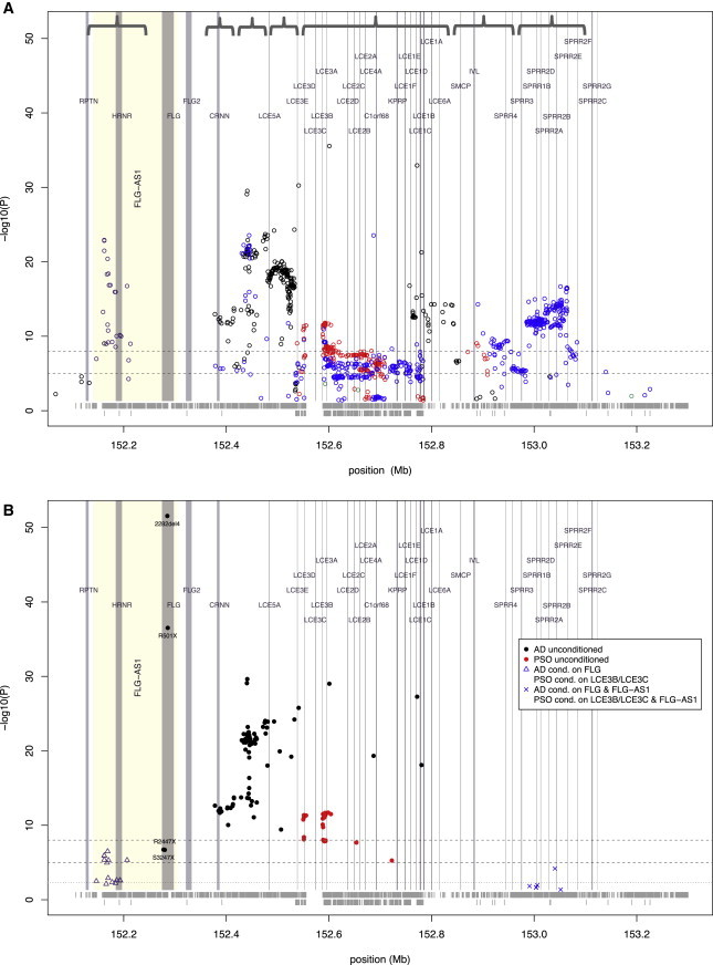
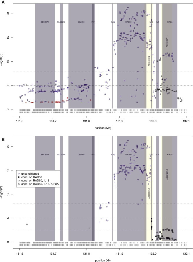
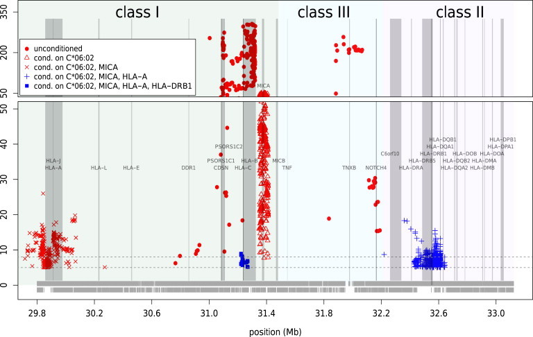
Atopic dermatitis and psoriasis are the two most common immune-mediated inflammatory disorders affecting the skin. Genome-wide studies demonstrate a high degree of genetic overlap, but these diseases have mutually exclusive clinical phenotypes and opposing immune mechanisms. Despite their prevalence, atopic dermatitis and psoriasis very rarely co-occur within one individual. By utilizing genome-wide association study and ImmunoChip data from >19,000 individuals and methodologies developed from meta-analysis, we have identified opposing risk alleles at shared loci as well as independent disease-specific loci within the epidermal differentiation complex (chromosome 1q21.3), the Th2 locus control region (chromosome 5q31.1), and the major histocompatibility complex (chromosome 6p21-22). We further identified previously unreported pleiotropic alleles with opposing effects on atopic dermatitis and psoriasis risk in PRKRA and ANXA6/TNIP1. In contrast, there was no evidence for shared loci with effects operating in the same direction on both diseases. Our results show that atopic dermatitis and psoriasis have distinct genetic mechanisms with opposing effects in shared pathways influencing epidermal differentiation and immune response. The statistical analysis methods developed in the conduct of this study have produced additional insight from previously published data sets. The approach is likely to be applicable to the investigation of the genetic basis of other complex traits with overlapping and distinct clinical features.


Figures

Figure 1
Figure 1
Study Design Abbreviations are as follows: CCMA, case control meta-analysis; MANTRA, meta-analysis of trans-ethnic association studies; BFD, Bayesian false discovery; PO, prior odds; conditional analysis for the MHC was also carried out with imputed classical HLA-allele (detailed in the Subjects and Methods).
Figure 2
Figure 2
Genome-wide Comparison of AD and Psoriasis (A) Mirrored Manhattan plots showing results of AD meta-GWAS (top) and psoriasis meta-GWAS (bottom). (B) Comparative analysis of AD and psoriasis in which SNVs are color coded to show AD-specific effect (black), psoriasis-specific effect (red), shared effects defined as alleles operating in the same direction (green), and opposing effects (blue). The genome-wide significance level is marked at p = 0.5 × 10−8 (Tmax = 6.0).
Figure 3
Figure 3
Regional Association within the Epidermal Differentiation Complex at 1q21.3 (A) Multinomial regression model with GWAS and ImmunoChip data. Seven blocks of linkage disequilibrium are indicated by curly brackets; black circles indicate AD-specific association, red circles indicate a psoriasis-specific association, blue circles represent opposing effects in AD and psoriasis, and green circles indicate shared effects. Vertical lines have been drawn to mark the positions of known genes and transcripts (identified from UCSC Genome Browser, GRCh37/hg19 accessed Feb. 2009) and the horizontal dotted lines indicate thresholds of suggestive and genome-wide significance (p = 10−5 and 10−8). The horizontal gray bands at the bottom indicate the coverage of the region by GWAS SNVs (upper row) and ImmunoChip SNVs (lower row). (B) Conditional regional association plot of stepwise logistic regression using GWAS and ImmunoChip data. The different colored symbols indicate association results after each step of analysis, as follows. Unconditioned results are shown by black dots to indicate association with AD and red dots to indicate association with psoriasis; blue triangles and blue crosses represent results after conditioning on the known disease-associated variants, FLG in AD and LCE3B-LCE3C deletion in psoriasis; SNVs indicated by the same symbol are in LD with the lead SNV of each stepwise conditional analysis (defined as r2 ≥ 0.5). Vertical lines are drawn to mark the positions of known genes and transcripts (identified from the UCSC Genome Browser GRCh37/hg19 accessed Feb. 2009), and horizontal dotted lines indicate significance thresholds of p = 0.005, 10−5, and 10−8. The horizontal gray bands at the bottom indicate the coverage of the region by GWAS SNVs (upper row) and ImmunoChip SNVs (lower row).
Figure 4
Figure 4
Regional Association within the Cytokine Cluster at 5q31.1 (A) Multinomial regression model with GWAS and ImmunoChip data. Black circles indicate AD-specific association, red circles indicate psoriasis-specific association, blue circles represent opposing effects in AD and psoriasis, and green circles indicate shared effects. Vertical gray shading marks the positions of known genes (identified from the UCSC Genome Browser GRCh37/hg19 accessed Feb. 2009), and horizontal dotted lines indicate suggestive and genome-wide significance thresholds (p = 10−5 and 10−8, respectively); results are shown for SNVs in LD with the lead SNV (defined as r2 ≥ 0.5). The horizontal bands at the bottom indicate the coverage of the region by GWAS SNVs (upper row) and ImmunoChip SNVs (lower row). (B) Conditional regional association plot of the EDC by multinomial regression of GWAS and ImmunoChip data. Different symbols indicate association results after each step of analysis, as follows. Unconditioned results are shown by blue circles representing opposing effects in AD and psoriasis; black dots show AD-specific association results after conditioning on the lead SNV in RAD50 (a gene reported to be associated with AD and psoriasis); black squares indicate the residual AD-specific association after conditioning on the lead SNVs in RAD50 and IL13 (genes reported to be associated with AD); and black triangles indicate the residual AD-specific association after additionally conditioning on the lead SNV in KIF3A (a gene reported to be associated with AD). SNVs indicated by the same symbol are in LD with the lead SNV of each stepwise conditional analysis (defined as r2 ≥ 0.5). Vertical gray shading marks the positions of known genes (identified from the UCSC Genome Browser GRCh37/hg19 accessed Feb. 2009), and horizontal dotted lines indicate significance thresholds of p = 0.005, 10−5, and 10−8; results are shown for SNVs in LD with the lead SNV (defined as r2 ≥ 0.5). The horizontal bands at the bottom indicate the coverage of the region by GWAS SNVs (upper row) and ImmunoChip SNVs (lower row).
Figure 5
Figure 5
Conditional Regional Association within the Major Histocompatibility Complex at 6p21–22 via GWAS and ImmunoChip Data Symbols indicate association results after each step of analysis, as follows. Unconditioned psoriasis-specific results are shown by red dots; red triangles show psoriasis-specific association results after conditioning on C06:02 (known to be strongly associated with psoriasis); red ×s indicate psoriasis-specific association after conditioning on C06:02 and MICA; blue +s indicate the association after conditioning on C06:02, MICA, and HLA-A with opposing effects on AD and psoriasis; and blue squares indicate the residual association after conditioning on C06:02, MICA, HLA-A, and HLA-DRB1 with opposing effects on AD and psoriasis. SNVs indicated by the same symbol are in LD with the lead SNV of each stepwise conditional analysis (defined as r2 ≥ 0.5). Vertical shading marks the positions of known genes (identified from the UCSC Genome Browser GRCh37/hg19 accessed Feb. 2009) and HLA classes; horizontal dotted lines indicate significance thresholds of p = 10−5 and p = 10−8; results are shown for SNVs in LD with the lead SNV (defined as r2 ≥ 0.5). The horizontal bands at the bottom indicate the coverage of the region by GWAS SNVs (upper row) and ImmunoChip SNVs (lower row).

References

    1. Deckert S., Kopkow C., Schmitt J. Nonallergic comorbidities of atopic eczema: an overview of systematic reviews. Allergy. 2014;69:37–45. - PubMed
    1. Griffiths C.E., Barker J.N. Pathogenesis and clinical features of psoriasis. Lancet. 2007;370:263–271. - PubMed
    1. Parisi R., Symmons D.P., Griffiths C.E., Ashcroft D.M., Identification and Management of Psoriasis and Associated ComorbidiTy (IMPACT) project team Global epidemiology of psoriasis: a systematic review of incidence and prevalence. J. Invest. Dermatol. 2013;133:377–385. - PubMed
    1. Deckers I.A., McLean S., Linssen S., Mommers M., van Schayck C.P., Sheikh A. Investigating international time trends in the incidence and prevalence of atopic eczema 1990-2010: a systematic review of epidemiological studies. PLoS ONE. 2012;7:e39803. - PMC - PubMed
    1. Henseler T., Christophers E. Disease concomitance in psoriasis. J. Am. Acad. Dermatol. 1995;32:982–986. - PubMed

Publication types