. 2015 Feb;125(2):652-64.
doi: 10.1172/JCI77701. Epub 2015 Jan 9.

HIF2α signaling inhibits adherens junctional disruption in acute lung injury

HIF2α signaling inhibits adherens junctional disruption in acute lung injury

Haixia Gong et al. J Clin Invest. 2015 Feb.

Erratum in

Abstract

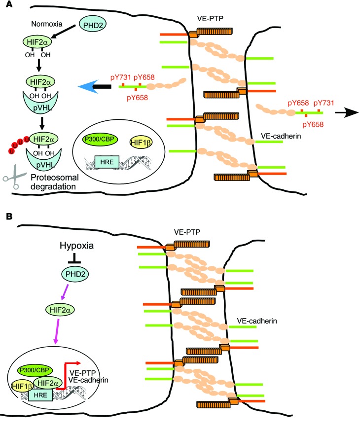
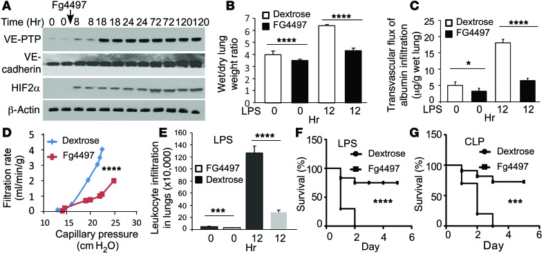
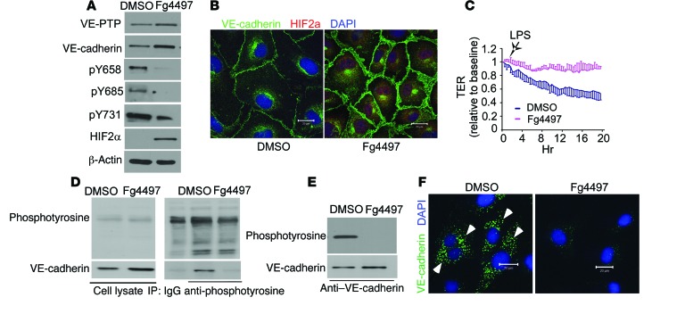
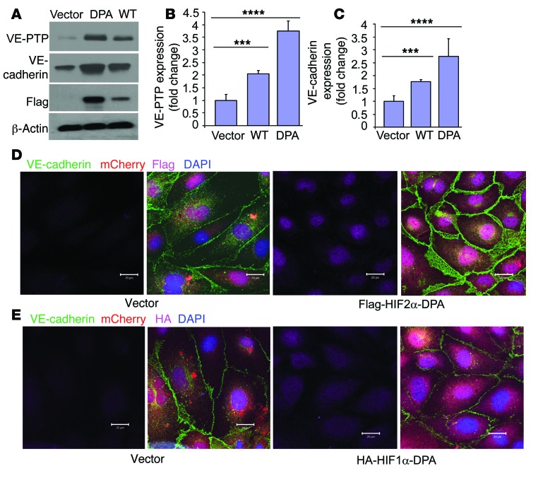
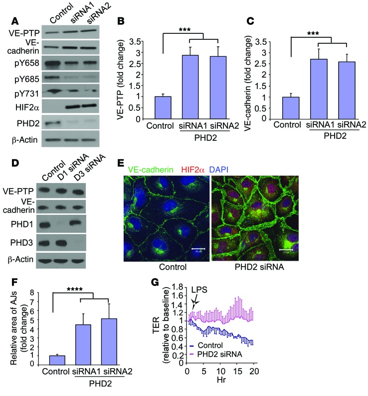
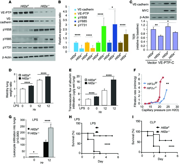
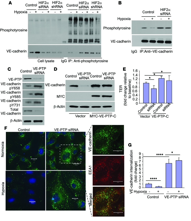
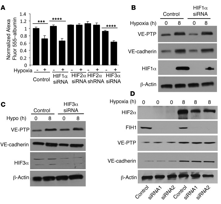
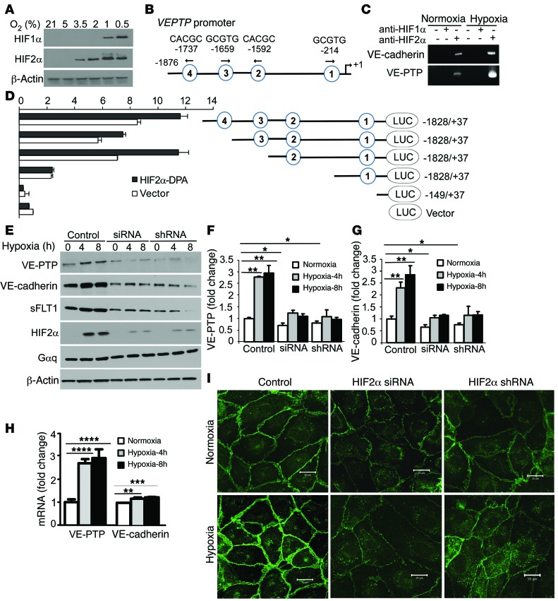
Vascular endothelial barrier dysfunction underlies diseases such as acute respiratory distress syndrome (ARDS), characterized by edema and inflammatory cell infiltration. The transcription factor HIF2α is highly expressed in vascular endothelial cells (ECs) and may regulate endothelial barrier function. Here, we analyzed promoter sequences of genes encoding proteins that regulate adherens junction (AJ) integrity and determined that vascular endothelial protein tyrosine phosphatase (VE-PTP) is a HIF2α target. HIF2α-induced VE-PTP expression enhanced dephosphorylation of VE-cadherin, which reduced VE-cadherin endocytosis and thereby augmented AJ integrity and endothelial barrier function. Mice harboring an EC-specific deletion of Hif2a exhibited decreased VE-PTP expression and increased VE-cadherin phosphorylation, resulting in defective AJs. Mice lacking HIF2α in ECs had increased lung vascular permeability and water content, both of which were further exacerbated by endotoxin-mediated injury. Treatment of these mice with Fg4497, a prolyl hydroxylase domain 2 (PHD2) inhibitor, activated HIF2α-mediated transcription in a hypoxia-independent manner. HIF2α activation increased VE-PTP expression, decreased VE-cadherin phosphorylation, promoted AJ integrity, and prevented the loss of endothelial barrier function. These findings demonstrate that HIF2α enhances endothelial barrier integrity, in part through VE-PTP expression and the resultant VE-cadherin dephosphorylation-mediated assembly of AJs. Moreover, activation of HIF2α/VE-PTP signaling via PHD2 inhibition has the potential to prevent the formation of leaky vessels and edema in inflammatory diseases such as ARDS.


Figures

Figure 9
Figure 9. Signaling pathways regulating HIF2α-induced VE-PTP expression.
(A) In normoxia, PHD2 hydroxylates HIF2α, resulting in its binding to pVHL, which targets HIF2α for proteasomal degradation. Basal levels of VE-PTP and VE-cadherin are expressed in ECs to maintain a restrictive endothelial barrier. VE-PTP–induced dephosphorylation of VE-cadherin maintains VE-cadherin at AJs and prevents VE-cadherin internalization. (B) In hypoxia, PHD2 activity is inhibited, and nonhydroxylated HIF2α accumulates in the nucleus and associates with constitutively expressed HIF1β and the coactivator CBP/P300 to transactivate VEPTP gene transcription through binding to HREs. VE-PTP interaction with VE-cadherin dephosphorylates VE-cadherin at Y658, Y685, and Y731 and inhibits VE-cadherin internalization, thus enhancing AJ assembly and endothelial barrier integrity.
Figure 8
Figure 8. PHD2 inhibition improves lung fluid balance and mortality.
(A) C57/BL6 mice were challenged with 5% dextrose or 25 mg/kg Fg4497 (same dose thereafter). WB was used to determine expression of the indicated proteins in lungs. (B and C) Three days after receiving Fg4497, C57/BL6 mice were challenged with PBS or 15 mg/kg LPS. Wet-to-dry lung weight ratios (B) and pulmonary transvascular albumin permeability (C) were measured. n = 5/group. (D) C57/BL6 mice received dextrose or Fg4497, and pulmonary transvascular fluid filtration was measured 3 days later. (E) Quantification of leukocytes in BAL fluid from Fg4497-treated mice 12 hours after 15 mg/kg LPS challenge. n = 4/group. ****P < 0.001 by Student’s t test (BE). (F and G) C57/BL6 mice were challenged with 25 mg/kg LPS (F) or subjected to CLP (G), and then 2 hours later received Fg4497. Survival rates were assessed by log-rank test. ***P < 0.005; ****P < 0.001. n = 10/group. Blot images were derived from samples run on parallel gels.
Figure 7
Figure 7. PHD2 inhibition promotes endothelial barrier integrity in vitro.
(A and B) HLMVECs were treated with 10 μM Fg4497 or DMSO for 8 hours. WB (A) or immunostaining (B) was performed to determine expression levels of the specified proteins. (C) HLMVECs pretreated with DMSO or Fg4497 were challenged with 1 μg/ml LPS, and AJ integrity was monitored by TER (n = 3/group). (D and E) Lysates of HLMVECs pretreated with DMSO or Fg4497 were precipitated with anti-phosphotyrosine (D) or anti–VE-cadherin (E) antibodies and detected by WB. (F) DMSO- or Fg4497-pretreated HLMVECs were labeled with anti–VE-cadherin antibody in the presence of 100 μM chloroquine. Internalization of VE-cadherin (arrowheads) was examined by confocal microscopy. Scale bars: 20 μm. Blot images were derived from samples run on parallel gels.
Figure 6
Figure 6. Expression of hydroxylation- and degradation-resistant HIF2α-mutant HIF2α-DPA in HLMVECs stabilizes the endothelial barrier via VE-PTP expression.
(A) HLMVECs were infected with lentivirus expressing a Flag-tagged WT HIF2α or with a hydroxylation- and degradation-resistant HIF2α-DPA mutant. VE-PTP and VE-cadherin protein expression was determined by WB. Quantification of VE-PTP (B) and VE-cadherin (C) expression (n = 3/group). (D and E) HLMVECs infected with lentivirus expressing WT, a HA-HIF1α-DPA mutant, or a Flag-HIF2α-DPA mutant were immunostained for VE-cadherin and analyzed by confocal microscopy. Scale bars: 20 μm. Blot images were derived from samples run on parallel gels. ***P < 0.005, ****P < 0.001 by Student’s t test.
Figure 5
Figure 5. PHD2 depletion enhances the integrity of the endothelial barrier and increases VE-PTP expression.
(A) HLMVECs were infected with control or lentiviral PHD2 siRNA, and WB was performed to determine the expression of VE-cadherin, VE-PTP, PHD2, and HIF2α as well as the extent of VE-cadherin tyrosine phosphorylation at Y658, Y685, and Y731. (B and C) Quantification of VE-PTP (B) and VE-cadherin (C) protein expression following PHD2 depletion (n = 3). (D) HLMVECs were infected with control or lentiviral PHD1 and PHD3 siRNAs, and WBs were used to compare expression levels of VE-cadherin, VE-PTP, PHD, and PHD3. (E) HLMVECs infected with control or lentiviral PHD2 siRNA were immunostained and analyzed by confocal microscopy. (F) Quantification of AJ thickness in random fields (n = 25–35 cells). (G) Serum-starved confluent HLMVECs infected with control or lentiviral PHD2 siRNA were challenged with 1 μg/ml LPS, and TER was monitored to determine AJ disassembly. TER values of each monolayer were normalized to baseline values (n = 4/group). ***P < 0.005, ****P < 0.001 by Student’s t test.
Figure 4
Figure 4. Endothelial-specific Hif2a deletion in mice increases lung vascular permeability through suppression of VE-PTP expression and VE-cadherin phosphorylation.
(A) Dissected lungs of Hif2afl/fl and Hif2aEC–/– mice were homogenized in RIPA buffer, and WB was performed to determine expression of the indicated proteins. (B) Quantification of VE-PTP and VE-cadherin expression and VE-cadherin phosphorylation (n = 4). (C) PMECs isolated from Hif2afl/fl and Hif2aEC–/– mice were transduced with control or MYC-VE-PTP-C lentivirus. Expression of MYC-VE-VTP-C and VE-cadherin was assessed by WB. Permeability of the endothelial monolayer was measured by TER assay under baseline normoxic conditions. n = 3–4. (D) Hif2afl/fl and Hif2aEC–/– mice were challenged with PBS or 15 mg/kg LPS i.p. Wet and dry lung weights were recorded to determine edema. n = 5 /group. (E) Hif2afl/fl and Hif2aEC–/– mice were injected with PBS or 15 mg/kg LPS. Pulmonary transvascular permeability to EBA was measured by detecting OD620 and OD740 of the formamide extract. Data represent the mean ± SD. n = 5/group. (F) Lungs of Hif2afl/fl and Hif2aEC–/– mice were isolated, and pulmonary transvascular filtration was assessed. Each plotted point represents a measurement from a different lung preparation. (G) Quantification of leukocytes in the BAL fluid from Hif2afl/fl and Hif2aEC–/– mice challenged with 15 mg/kg LPS. n = 4/group. (BG) *P < 0.05, **P < 0.01, ****P < 0.001 by Student’s t test. (H and I) Hif2afl/fl and Hif2aEC–/– mice were challenged with 25 mg/kg LPS i.p. (H) or polymicrobial sepsis was induced by CLP (I), and survival was monitored. n = 10/group. Differences in mortality were analyzed by log-rank test. Blot images were derived from samples run on parallel gels. ****P < 0.001 by log-rank test.
Figure 3
Figure 3. HIF2α-induced VE-PTP expression mediates AJ integrity through retention of VE-cadherin at AJs.
(A and B) HLMVECs were exposed to normoxia or 1% O2 for 8 hours and harvested in modified RIPA buffer. Cell lysates were precipitated with anti-phosphotyrosine antibody (A) or anti–VE-cadherin antibody (B), and IPs were then probed by WB using the converse antibody. (C) HLMVECs were infected with control or VE-PTP siRNA lentivirus. Expression of VE-VTP and VE-cadherin and phosphorylation of the indicated tyrosines in VE-cadherin were assessed by WB. (D) Control and VE-PTP siRNA lentivirus-infected HLMVECs were subjected to overexpression of an active MYC-VE-PTP-C fragment. Expression of VE-cadherin and MYC-VE-PTP-C was detected by WB. (E) Permeability of the endothelial monolayer of these cells used in D was measured by TER assay under basal conditions (n = 3–4). (F) Control and VE-PTP siRNA lentivirus–infected HLMVECs were exposed to normoxia or 1% O2 for 8 hours, labeled with anti–VE-cadherin antibody (BV9) in the presence of 100 μM chloroquine for 4 hours at 37°C to visualize internalized VE-cadherin in vesicles, and then subjected to a mild acid buffer wash to remove noninternalized VE-cadherin. Cells were fixed and subjected to immunostaining using anti-EEA1 antibody to visualize endosomes. Internalization of VE-cadherin was examined by confocal microscopy. Scale bars: 20 μm. (G) Quantification of internalized VE-cadherin puncta from 4 to 8 randomly chosen fields. Blot images were derived from samples run on parallel gels. *P < 0.05, ****P < 0.001 by Student’s t test.
Figure 2
Figure 2. Hypoxia-induced stabilization of the endothelial barrier is mediated by HIF2α.
(A) Control, HIF1α-, HIF2α-, or HIF3α-depleted HLMVECs were grown on Transwell plates and exposed to 1% O2 (hypoxia) for 8 hours with an Alexa Fluor 555–albumin tracer to assess endothelial barrier permeability. Tracer concentrations were determined in the lower-chamber media. ***P < 0.005, ****P < 0.001 by Student’s t test. (BD) HLMVECs infected with lentiviral control vector, HIF1α siRNA (B), HIF3α siRNA (C), or FIH1 siRNA (D) were exposed to normoxia or 1% O2. Expression of the indicated proteins was evaluated by WB. n = 3/group (AD). Blot images were derived from samples run on parallel gels.
Figure 1
Figure 1. HIF2α induces VE-PTP expression and enhances the integrity of endothelial AJs.
(A) HLMVECs were exposed to varying concentrations of O2 for 8 hours. Expression of HIF1α and HIF2α was assessed by WB. (B) Representation of the VEPTP promoter region. HREs are shown by circled numbers, and their sequences are displayed. (C) HLMVECs were exposed to normoxia or 1% O2 for 8 hours. A ChIP assay was performed to amplify the VEPTP and VE-cadherin promoters. (D) 293T cells were transfected with an HIF2α-DPA expression plasmid containing luciferase reporter constructs. Luciferase values were normalized to β-gal values. A schematic representation of corresponding deletion constructs is presented in the right panel. (E) HLMVECs infected with lentiviral HIF2α siRNA/shRNA were exposed to normoxia or 1% O2. Expression of VE-cadherin, VE-PTP, and HIF2α was assessed by WB. (F-G) Quantification of VE-PTP (F) and VE-cadherin (G) protein levels. (H) HLMVECs were exposed to normoxia or 1% O2. Expression of VEPTP and VE-cadherin at the mRNA level was assessed by quantitative PCR. (I) AJ integrity of HLMVECs was examined by VE-cadherin immunostaining using confocal microscopy. Scale bars: 20 μm. n = 3/group (FH). Blot images were derived from samples run on parallel gels. *P < 0.05; **P < 0.01; ***P < 0.005; ****P < 0.001 by Student’s t test.

Similar articles

Cited by

References

    1. Mehta D, Malik AB. Signaling mechanisms regulating endothelial permeability. Physiol Rev. 2006;86(1):279–367. doi: 10.1152/physrev.00012.2005. - DOI - PubMed
    1. Kuppers V, Vockel M, Nottebaum AF, Vestweber D. Phosphatases and kinases as regulators of the endothelial barrier function. Cell Tissue Res. 2014;355(3):577–586. doi: 10.1007/s00441-014-1812-1. - DOI - PubMed
    1. Semenza GL. Targeting HIF-1 for cancer therapy. Nat Rev Cancer. 2003;3(10):721–732. doi: 10.1038/nrc1187. - DOI - PubMed
    1. Farahani RM, Sarrafpour B, Simonian M, Li Q, Hunter N. Directed glia-assisted angiogenesis in a mature neurosensory structure: pericytes mediate an adaptive response in human dental pulp that maintains blood-barrier function. J Comp Neurol. 2012;520(17):3803–3826. doi: 10.1002/cne.23162. - DOI - PubMed
    1. Appelhoff RJ, et al. Differential function of the prolyl hydroxylases PHD1, PHD2, and PHD3 in the regulation of hypoxia-inducible factor. J Biol Chem. 2004;279(37):38458–38465. doi: 10.1074/jbc.M406026200. - DOI - PubMed

Publication types

MeSH terms

Substances