Clinical Trial

Global transcriptional disturbances underlie Cornelia de Lange syndrome and related phenotypes

Bo Yuan et al. J Clin Invest. 2015 Feb.

Abstract

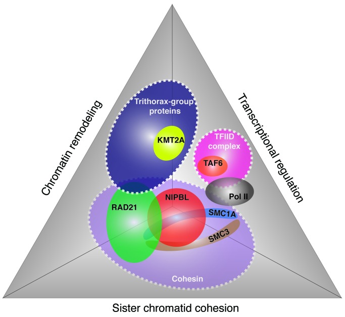
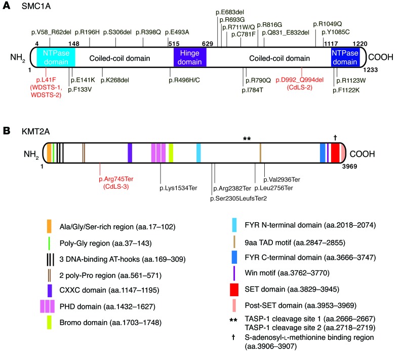
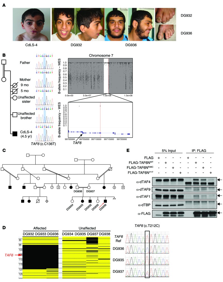
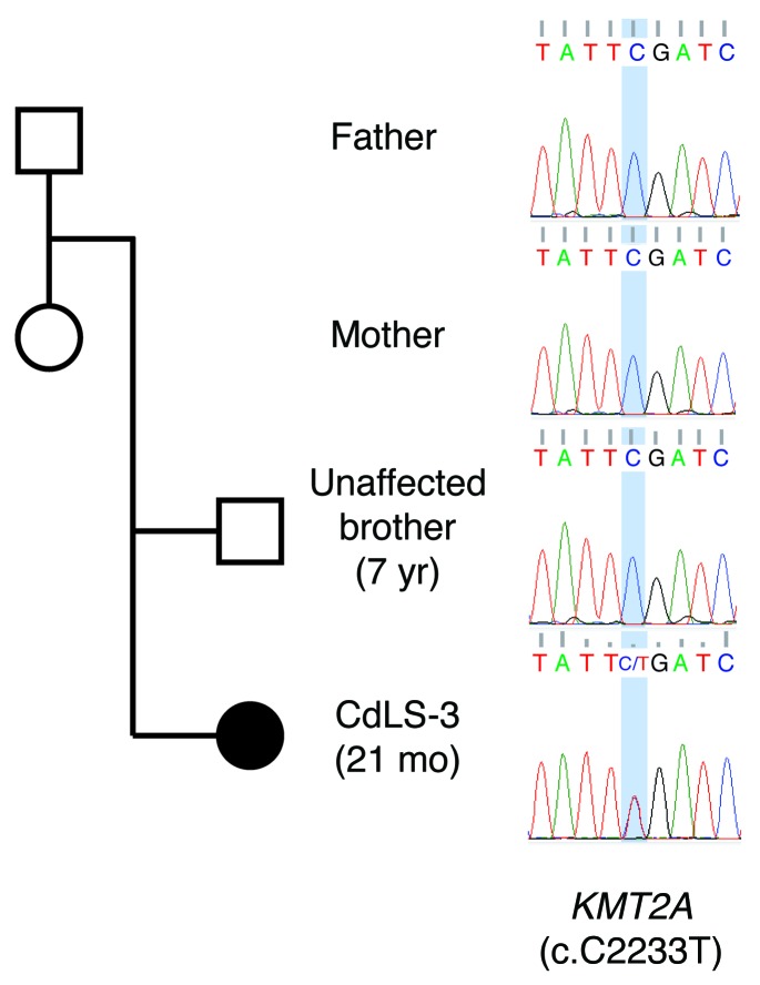
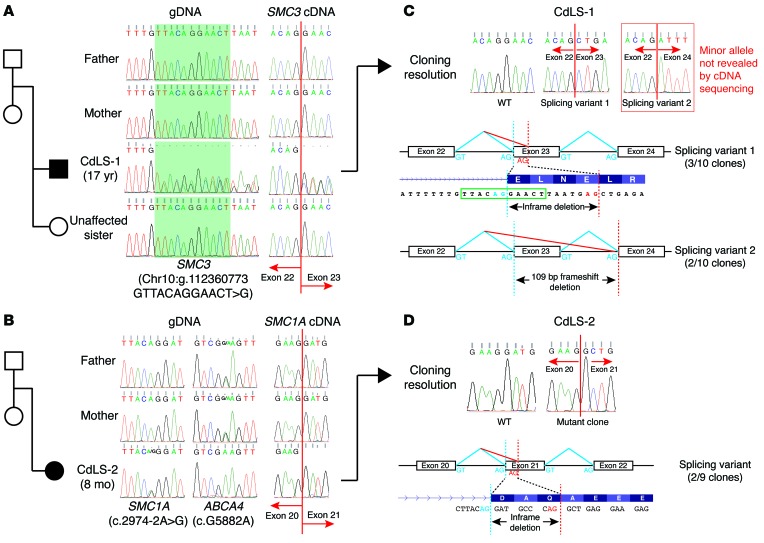
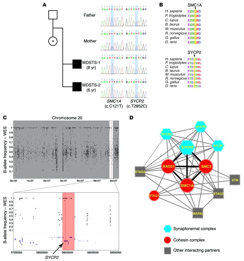
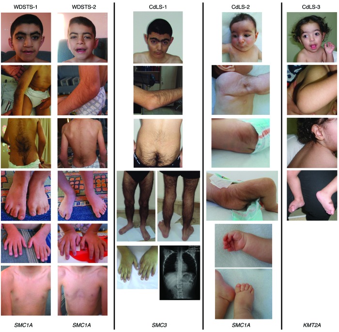
Cornelia de Lange syndrome (CdLS) is a genetically heterogeneous disorder that presents with extensive phenotypic variability, including facial dysmorphism, developmental delay/intellectual disability (DD/ID), abnormal extremities, and hirsutism. About 65% of patients harbor mutations in genes that encode subunits or regulators of the cohesin complex, including NIPBL, SMC1A, SMC3, RAD21, and HDAC8. Wiedemann-Steiner syndrome (WDSTS), which shares CdLS phenotypic features, is caused by mutations in lysine-specific methyltransferase 2A (KMT2A). Here, we performed whole-exome sequencing (WES) of 2 male siblings clinically diagnosed with WDSTS; this revealed a hemizygous, missense mutation in SMC1A that was predicted to be deleterious. Extensive clinical evaluation and WES of 32 Turkish patients clinically diagnosed with CdLS revealed the presence of a de novo heterozygous nonsense KMT2A mutation in 1 patient without characteristic WDSTS features. We also identified de novo heterozygous mutations in SMC3 or SMC1A that affected RNA splicing in 2 independent patients with combined CdLS and WDSTS features. Furthermore, in families from 2 separate world populations segregating an autosomal-recessive disorder with CdLS-like features, we identified homozygous mutations in TAF6, which encodes a core transcriptional regulatory pathway component. Together, our data, along with recent transcriptome studies, suggest that CdLS and related phenotypes may be "transcriptomopathies" rather than cohesinopathies.


Figures

Figure 7
Figure 7. Diagram summarizing interaction network potentially encompassing all the proteins encoded by the genes identified in the study.
Three categories of cellular functions (sister chromatid cohesion, chromatin remodeling, and transcriptional regulation) are shown. The proteins are illustrated in colored spheres. NIPBL, SMC1A, SMC3, and RAD21 are the 4 components in cohesin, which is involved in sister chromatid cohesion, chromatin remodeling, and transcriptional regulation. The interaction between trithorax-group proteins and the RAD21 links KMT2A into this interaction network. RNA Pol II interacts with both TFIID and cohesin and then potentially links TAF6, a core component of TFIID complex, to the cohesin.
Figure 6
Figure 6. Summaries of the damaging, potentially disease-associated mutations identified in this study and those reported previously.
(A) Summary of SMC1A mutations reported in patients with CdLS (in black) and mutations reported in this study (in red). The domains of SMC1A are illustrated and annotated in the diagram. (B) Summary of KMT2A mutations reported in patients with WDSTS (in black) and the mutation reported in this study (in red). The domains of KMT2A are illustrated, with each annotated underneath the diagram.
Figure 5
Figure 5. TAF6 variants identified in the Turkish family of CdLS-4 and the Saudi family.
(A) Patient photographs showing the clinical features of CdLS-4 and 2 patients (DG932 and DG936) from the Saudi family. (B) The pedigree of the CdLS-4 family and the segregation of the TAF6 variant. B-allele frequency plots from the WES data for CdLS-4 are shown on the right. The region of AOH is shown as a white region between the flanking gray areas. Top panel: B-allele frequency plot of the entire chromosome 7; bottom panel: zoomed-in view of the region encompassing TAF6 indicated by the black arrow. (C) The pedigree of the Saudi family. Red arrow indicates the patient that underwent WES. (D) The homozygosity mapping on the left shows the region of AOH (black blocks) encompassing TAF6 (indicated by red arrow) in DG932, DG933 and DG936. The Sanger-sequencing chromatograms on the right show homozygous WT in the control and DG935, homozygous mutation in DG936, and heterozygous mutation in the mother, DG937. (E) Co-IP in Drosophila S2 cells testing TAF6-binding affinity. S2 cells were treated with dsRNA targeting dTAF6C. FLAG-TAF6NWT, FLAG-TAF6NR46C, and FLAG-TAF6NI71T correspond to the S2 cells transfected with WT or mutated dTAF6N as indicated. FLAG, negative control. Four TFIID components (dTAF1, dTAF4, dTAF9, and dTBP) were detected with Western blot using antibodies against each indicated component. Left 4 columns, input; right 4 columns, proteins co-IP with FLAG-TAF6N. The lanes were run on the same gel, but were noncontiguous.
Figure 4
Figure 4. Segregation analysis of the KMT2A mutation in the family of CdLS-3.
The gene and nt changes are shown underneath the Sanger-sequencing chromatograms.
Figure 3
Figure 3. Variants in SMC3 and SMC1A identified in patients with CdLS (CdLS-1 and CdLS-2).
(A and B) Pedigree of the families of CdLS-1 and CdLS-2 and their corresponding Sanger-sequencing chromatograms showing the segregation analyses. The chromatograms on the left show the segregation of the variants in each family. The gene and nt changes are shown underneath the chromatograms. The chromatograms on the right show the outcomes of alternative splicing introduced by the splicing-site variants and their segregations in the family. Red lines in the middle split the 2 adjacent exons. (C and D) Cloning method resolves the compound alleles in patient-derived cDNA. Different kinds of alternative spliced alleles are shown in the top panel in forms of Sanger-sequencing chromatograms. The outcomes of alternative splicing are shown in the lower panels. Blue dashed lines, site of normal splicing acceptor; red dashed lines, site of alternative splicing acceptors.
Figure 2
Figure 2. The variants in SMC1A and SYCP2 identified in the patients with WDSTS (WDSTS-1 and WDSTS-2).
(A) Pedigree of the family and the Sanger-sequencing chromatogram showing the segregation analysis of variants identified in SMC1A and SYCP2. The blue shading in the chromatograms marks the position of the variants. The genes and nt changes are shown underneath the chromatograms. (B) Peptide alignments showing the conservation of the affected aa across different species. First panel: Leu41 in SMC1A. Second panel: Ile951 in SYCP2. All these aa are highlighted by yellow shading, and they are highly conserved across the species. (C) The B-allele frequency plots from the WES data of WDSTS-1. The upper panel shows the overall B-allele frequencies of the entire chromosome 20. The lower panel shows the zoomed-in view of the region surrounding the variant (Chr20: g.58455447 A>G [hg19]). The region highlighted by red shading represents the AOH region, including the gene SYCP2, which is indicated by the black arrow. (D) The interaction network that includes both SC and the cohesin complex. Blue hexagons, major components of the SC; red circles, major components of the cohesin complex; gray squares, other interacting partners involved in this network. Heavy black lines show the strong interactions between the SYCP2 and the 3 major components of the cohesin complex: SMC1A, SMC3, and RAD21.
Figure 1
Figure 1. Photographs showing the representative clinical features of the patients WDSTS-1, WDSTS-2, CdLS-1, CdLS-2, and CdLS-3.
Note the eyebrows of the WDSTS-1 and WDSTS-2 are bushy, whereas the eyebrows of CdLS-1 and CdLS-3 have the typical arched appearance of CdLS with synophrys. WDSTS-1 and WDSTS-2 also have square and bulbous nasal tips. The second row of images demonstrates hirsutism of elbows in WDSTS-1, WDSTS-2, CdLS-1, and CdLS-2. Also of note is hirsutism of lower back, buttocks, legs, and face in the subjects. All individuals have modest brachydactyly of fingers and/or toes, and WDSTS-1, WDSTS-2, and CdLS-1 have fifth finger clinodactyly. WDSTS-1 and WDSTS-2 have pectus excavatum, and in CdLS-1, mild mid-thoracic scoliosis can be seen on the plain radiograph of the chest.

References

    1. Brachmann W. Ein fall von symmetrischer monodaktylie durch Ulnadefekt, mit symmetrischer flughautbildung in den ellenbeugen, sowie anderen abnormitaten (zwerghaftogkeit, halsrippen, behaarung). Jarb Kinder Phys Erzie. 1916;84:225–235.
    1. de Lange C. Sur un type nouveau de degenerescence (typus Amstelodamensis). Arch Med Enfants. 1933;36:713–719.
    1. Tonkin ET, Wang TJ, Lisgo S, Bamshad MJ, Strachan T. NIPBL, encoding a homolog of fungal Scc2-type sister chromatid cohesion proteins and fly Nipped-B, is mutated in Cornelia de Lange syndrome. Nat Genet. 2004;36(6):636–641. doi: 10.1038/ng1363. - DOI - PubMed
    1. Krantz ID, et al. Cornelia de Lange syndrome is caused by mutations in NIPBL, the human homolog of Drosophila melanogaster Nipped-B. Nat Genet. 2004;36(6):631–635. doi: 10.1038/ng1364. - DOI - PMC - PubMed
    1. Musio A, et al. X-linked Cornelia de Lange syndrome owing to SMC1L1 mutations. Nat Genet. 2006;38(5):528–530. doi: 10.1038/ng1779. - DOI - PubMed

Publication types

MeSH terms