. 2015 Jan 9:6:5614.
doi: 10.1038/ncomms6614.

Mutations in PNPLA6 are linked to photoreceptor degeneration and various forms of childhood blindness

Collaborators, Affiliations

Mutations in PNPLA6 are linked to photoreceptor degeneration and various forms of childhood blindness

S Kmoch et al. Nat Commun. .

Abstract

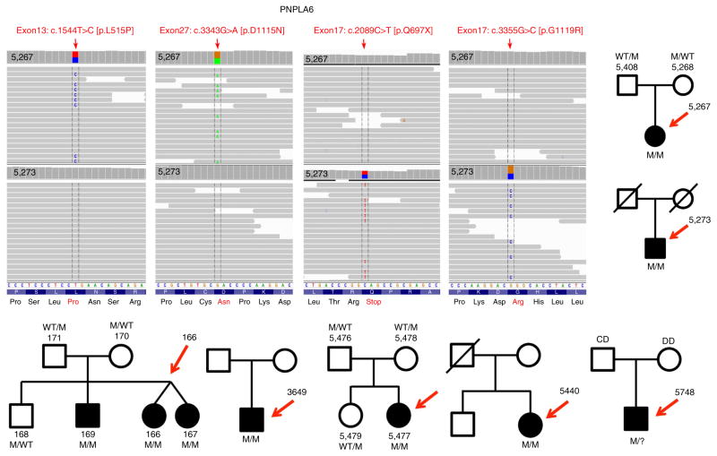
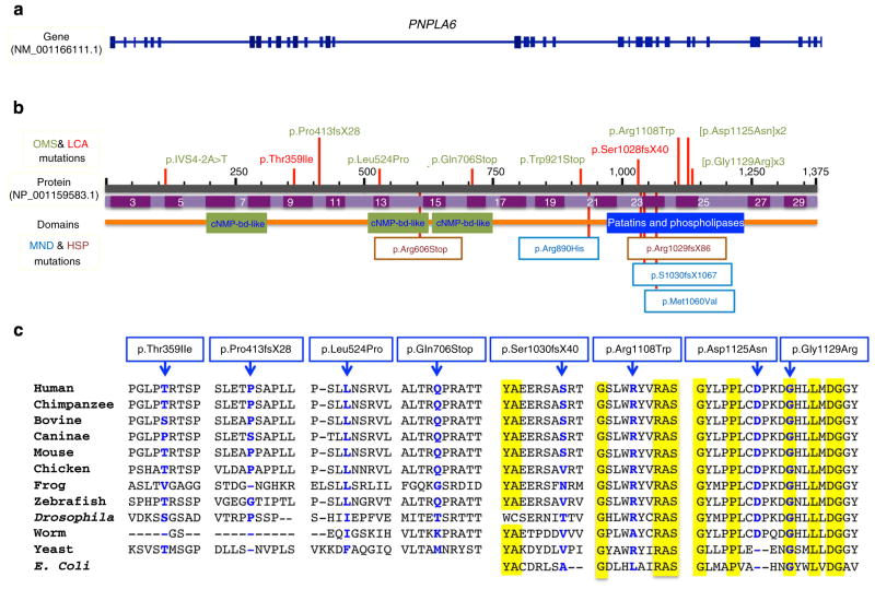
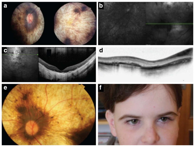
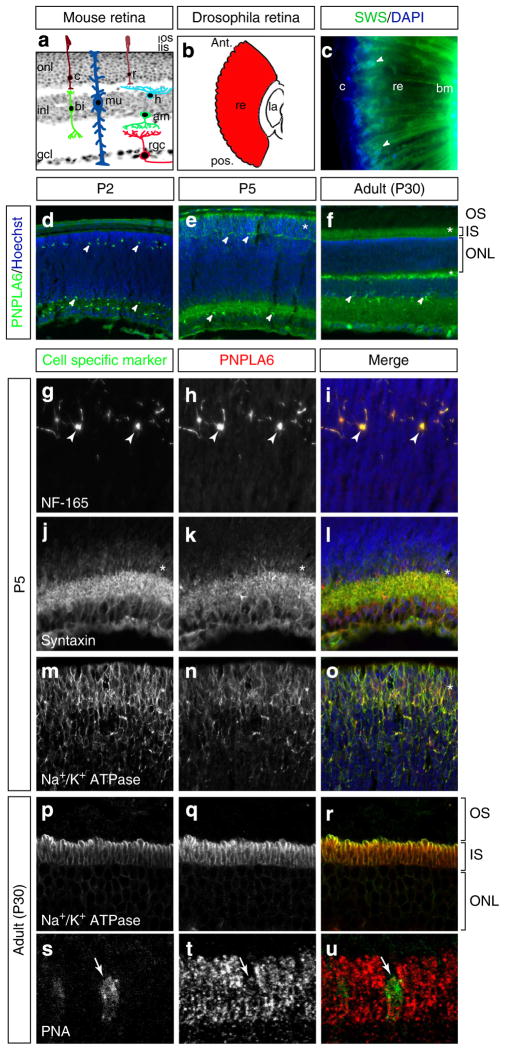
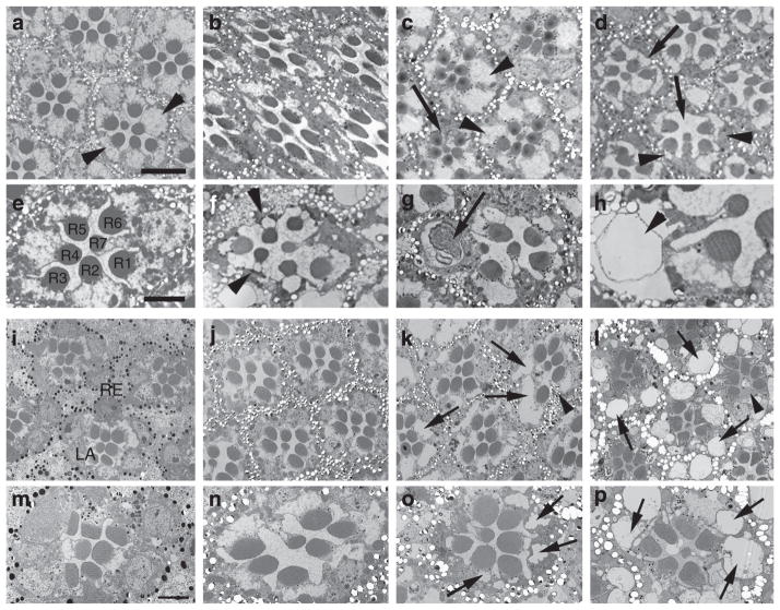
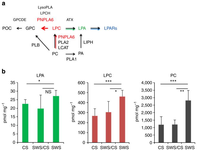
Blindness due to retinal degeneration affects millions of people worldwide, but many disease-causing mutations remain unknown. PNPLA6 encodes the patatin-like phospholipase domain containing protein 6, also known as neuropathy target esterase (NTE), which is the target of toxic organophosphates that induce human paralysis due to severe axonopathy of large neurons. Mutations in PNPLA6 also cause human spastic paraplegia characterized by motor neuron degeneration. Here we identify PNPLA6 mutations in childhood blindness in seven families with retinal degeneration, including Leber congenital amaurosis and Oliver McFarlane syndrome. PNPLA6 localizes mostly at the inner segment plasma membrane in photoreceptors and mutations in Drosophila PNPLA6 lead to photoreceptor cell death. We also report that lysophosphatidylcholine and lysophosphatidic acid levels are elevated in mutant Drosophila. These findings show a role for PNPLA6 in photoreceptor survival and identify phospholipid metabolism as a potential therapeutic target for some forms of blindness.


Conflict of interest statement

Competing financial interests: The authors declare no competing financial interests.

Figures

Figure 1
Figure 1. PNPLA6 mutations in individuals from seven families with childhood blindness
Sequencing reads are aligned against positive strand of hg19. Vertical arrows represent the 100 paired-end reads in 5,267 and 5,273 patients, respectively. Both patients carry heterozygous mutations in PNPLA6: 5,267 has 2 missense mutations c.1571T>C (leucine to proline substitution at codon 524) and c.3373G>A (aspartic acid to asparagine at codon 1125). Patient 5,273 has a nonsense mutation c.2116C>T, which result in a glutamine to stop codon at codon 706, leading to loss of the last 669 amino acids of the protein and a missense mutation c.3385G>C glycine to arginine at codon 1129. All of the complementary DNA positions are based on PNPLA6 NM_001166111.1. Moreover, shown are seven families/pedigrees with various forms of childhood blindness due to photoreceptor degeneration in OMS and LCA. Chromatograms of the PNPLA6 mutations, pedigrees and co-segregation of the mutations are shown for each family (Supplementary Fig. 1).
Figure 2
Figure 2. Mutations identified in PNPLA6 patients
Shown here is the genetic structure (a) and the protein structure (b) with all the mutations (ten) identified in the current study indicated above the PNPLA6 protein. Previously reported mutations causing the paraplegia syndrome (SPG39) are shown below the protein. A line-up of the affected protein regions from various species (from E. coli to man) is shown in c, showing the marked and significant evolutionary conservation of the residues, some down to E. coli.
Figure 3
Figure 3. Retinal phenotypes of children with PNPLA6 mutations
(a) A retinal photo shows severe choroidal and retinal atrophy ‘choroideremia like’ in patient 5,267. (b) Fundus autofluorescence (FAF) shows severely abnormal and grossly absent lipofuscin metabolism. (c) Optical coherence tomography reveals severe photoreceptor loss, retinal thinning and retinal remodelling. (d) Using optical coherence tomography, we found that the inner retinal layers are also abnormal. (e) Another child (167) with mutations in PNPLA6 shows a strikingly similar retinal appearance as shown in a, again illustrating the ‘choroideremia-like’ retinal changes. (f) Full-face photo of a child with PNPLA6 mutations, illustrating the extremely long lashes.
Figure 4
Figure 4. PNPLA6 expression in the mouse and Drosophila retina
(a) Section of a mouse retina superimposed with illustrations of the cell types found in the three different layers. onl, outer nuclear layer; inl, inner nuclear layer; gcl, ganglion cell layer; os, outer segment; is, inner segment; r, rod; c, cone; mu, Müller cell; rgc, retinal ganglion cell; bi, bipolar cell; h, horizontal cell; am, amacrine cell. (b) Illustration of a Drosophila retina along the longitudinal axis. ant, anterior; pos, posterior; re, retina; la, lamina. (c) Immunostaining for SWS in longitudinal sections of fly retina. SWS is expressed surrounding the nuclei and along the entire length of the photoreceptor (arrowheads). bm, basement membrane; c, cornea; re, retina. (df) Time course of PNPLA6 expression in the mouse retina, as indicated. PNPLA6 is expressed in cells of the inner and outer retina at all stages examined (arrowheads), as well as in the photoreceptor inner segment area (asterisk). Co-immunostaining of mouse retinal sections at P5 with PNPLA6 and the horizontal cell marker neurofilament-165 (NF-165; gi), amacrine cell marker syntaxin (jl) and the photoreceptor cell marker Na+/K+-ATPase (mo). Arrowheads point to positive cells and asterisks point to area of positive staining. (pr) Confocal z-stack projection of an adult mouse retinal sections (P30) co-immunostained with PNPLA6 and the photoreceptor inner segment plasma membrane marker Na+/K+-ATPase. PNPLA6 localizes mostly to the plasma membrane, although some staining is detected inside the inner segment. (su) Co-immunostaining of PNPLA6 with the cone photoreceptor marker peanut agglutinin (PNA) showing expression of PNPLA6 in cones (arrow). Sections were counterstained with Hoechst (blue) to reveal nuclei.
Figure 5
Figure 5. Loss of SWS causes photoreceptor degeneration in Drosophila
Electron microscopy studies from a 3-week-old wild-type fly show intact ommatidia with seven photoreceptor cells (R1–7) present (a,e; arrowheads in a point to the photoreceptor cell somata). Performing sections from the retina of a sws1 mutant fly shortly before eclosion (pharate adult) shows that photoreceptors develop normally (b) and persist during the first 2 weeks of adulthood (c, 14 days), although occasionally ommatidia missing a single photoreceptor can be found (arrow). However, after 23 days of ageing (which, due to the reduced live span of sws1, is shortly before these flies die), photoreceptor degeneration is evident in all ommatidia by the loss of photoreceptors (arrows in d), and in the condensed and darkly stained cell somata of the photoreceptors that are still present (arrowheads). In addition, vacuoles can be found in the somata (arrowheads in f,h) and the rhabdomeres are shrunken. In some photoreceptors, membranous structures can be found (arrow in g). Knocking down SWS specifically in the eye using an RNA interference (RNAi) construct expressed by GMR-GAL4 showed that newly enclosed flies look normal with all the seven photoreceptors R1–7 present (i,m). At 7 days of age the ommatidia still look wild type (j,n), but at 4 weeks the photoreceptors clearly show degeneration with vacuoles forming in the photoreceptor somata (arrows in k,o) and loss of some photoreceptors (arrowhead in k). Raising the flies in the dark resulted in a comparable photoreceptor degeneration in 30-day-old flies (l,p). Scale bar, 4 μm (a), 2 μm (e).
Figure 6
Figure 6. Phospholipid metabolism is altered in SWS mutant flies
(a) PNPLA6 (NTE)-related metabolic pathway. POC, phosphorylcholine; GPC, glycerolphosphocholine; LPC, lysophosphatidylcholine; LPA, lysophosphatidic acid; LPARs, lysophosphatidic acid receptors; PCH, phosphatidylcholine; PA, phosphatidic acid; GPCDE, glycerolphosphocholinediesterase; NTE, neuropathy target esterase; LCAT, lecithin-cholesterol acyltransferase; PLA1, phospholipase A1; PLA2, phospholipase A1; PLB, phospholipase B; LPCH, lysophosphatidylcholine hydrolase; LIPH, lipase H; ATX, autotaxin. (b) Elevated PC, LPC and LPA levels in SWS mutant flies. Wild-type Drosophila (CS), (n = 8); mutant heterozygotes (SWS/CS), n = 4; sws1-null mutants (SWS), n = 5. PC, LPC and LPA levels (y axis) are expressed in pmol of individual compounds per mg of protein. The means±s.d. for individual groups and P-values (t-test) are indicated; *P ≤ 0.05, **P ≤ 0.01, ***P ≤ 0.001; n.s., not significant. Experiments were repeated twice.

References

    1. Dudek BR, Richardson RJ. Evidence for the existence of neurotoxic esterase in neural and lymphatic tissue of the adult hen. Biochem Pharmacol. 1982;31:1117–1121. - PubMed
    1. Johnson MK. Initiation of organophosphate-induced delayed neuropathy. Neurobehav Toxicol Teratol. 1982;4:759–765. - PubMed
    1. Glynn P. Neuropathy target esterase and phospholipid deacylation. Biochim Biophys Acta. 2005;1736:87–93. - PubMed
    1. Johnson MK. The delayed neurotoxic effect of some organophosphorus compounds. Identification of the phosphorylation site as an esterase. Biochem J. 1969;114:711–717. - PMC - PubMed
    1. Glynn P. NTE: one target protein for different toxic syndromes with distinct mechanisms? Bioessays. 2003;25:742–745. - PubMed

Publication types

MeSH terms