. 2015 Mar:15:139-49.
doi: 10.1016/j.actbio.2014.12.027. Epub 2015 Jan 7.

Myoconductive and osteoinductive free-standing polysaccharide membranes

Affiliations

Myoconductive and osteoinductive free-standing polysaccharide membranes

Sofia G Caridade et al. Acta Biomater. 2015 Mar.

Abstract

Free-standing (FS) membranes have increasing applications in the biomedical field as drug delivery systems for wound healing and tissue engineering. Here, we studied the potential of free-standing membranes made by the layer-by-layer assembly of chitosan and alginate to be used as a simple biomimetic system of the periosteum. The design of a periosteum-like membrane implies the elaboration of a thick membrane suitable for both muscle and bone formation. Our aim was to produce well-defined ∼50 μm thick polysaccharide membranes that could be easily manipulated, were mechanically resistant, and would enable both myogenesis and osteogenesis in vitro and in vivo. The membranes were chemically crosslinked to improve their mechanical properties. Crosslinking chemistry was followed via Fourier transform infrared spectroscopy and the mechanical properties of the membranes were assessed using dynamic mechanical analysis. The loading and release of the potent osteoinductive growth factor bone morphogenetic protein 2 (BMP-2) inside and outside of the FS membrane was followed by fluorescence spectroscopy in a physiological buffer over 1 month. The myogenic and osteogenic potentials of the membranes in vitro were assessed using BMP-2-responsive skeletal myoblasts. Finally, their osteoinductive properties in vivo were studied in a preliminary experiment using a mouse ectopic model. Our results showed that the more crosslinked FS membranes enabled a more efficient myoblast differentiation in myotubes. In addition, we showed that a tunable amount of BMP-2 can be loaded into and subsequently released from the membranes, depending on the crosslinking degree and the initial BMP-2 concentration in solution. Only the more crosslinked membranes were found to be osteoinductive in vivo. These polysaccharide-based membranes have strong potential as a periosteum-mimetic scaffold for bone tissue regeneration.

Keywords: Biomaterials; Free-standing membranes; Layer-by-layer; Osteoinduction; Polysaccharides.


Figures

FIGURE 1
FIGURE 1. Optical microscopy and SEM images of free-standing (CHI/ALG)200 membranes
(A) Images of a dry (left) or hydrated (right) (CHI/ALG)200 free-standing membrane. Scale bar: 1 cm. (B) SEM observations of the upper side of the native (CHI/ALG)200 membrane and for the FS membranes crosslinked at increasing concentration of EDC from 10 to 50 mg/mL. Scale bar is 10 μm. (B’) Corresponding cross-sections of the crosslinked FS membranes. Scale bar: 20 μm.
FIGURE 2
FIGURE 2. Crosslinking and mechanical properties of the FS membranes
(A) FTIR spectra of native and crosslinked (CHI/ALG)200 FS membranes obtained at increasing EDC concentrations and (A’) differences between the spectra of the crosslinked FS membranes to that of the native membrane. (B, B’) Results of the DMA experiments performed at 37°C in PBS over 0.1 to 20 Hz. (B) Variation of the storage modulus (E’) and (B’) of the loss factor (tan δ).
FIGURE 3
FIGURE 3. C2C12 myoblast proliferation and differentiation on the FS polysaccharide membranes
Cells were cultured on the FS membranes crosslinked at EDC10, EDC30 and EDC50. (A) CLSM images of C2C12 myoblasts were taken after 24 h in GM and after 5 days in DM. The actin cytoskeleton was stained with rhodamine-phalloidin, the nuclei were stained with DAPI and the myotubes with troponin T. Scale bar is 200 μm. (B) % of proliferating cells measured by the EdU assay after 24 h in GM and (C) Quantification of the fusion index after 5 days in DM on EDC10, EDC30 and EDC50 FS membranes (mean ± SEM of three independent experiments, * p < 0.05).
FIGURE 4
FIGURE 4
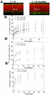
Quantification of BMP-2 loaded in and released from the (CHI/ALG) FS membranes. (A) CLSM images of the EDC50 FS membrane labeled with Alexa 568 (red) and loaded with BMP-2 CF (green). Two BMP-2 layers on the lower and upper side of the membrane were observed over a period of 8 months. (B) Release profiles of the EDC10, EDC30, and EDC50 FS membranes over a period of one month for an initial BMP-2 loading concentration of 20 μg/ml; (B’, B”) Release profiles of the EDC10 and EDC50 FS membranes over a period of one month for an initial BMP-2 loading concentration of 60 μg/ml (B’) and 100 μg/ml (B”). (mean ± SEM of three samples).
FIGURE 5
FIGURE 5
Proliferation and osteogenic differentiation of C2C12 myoblasts on BMP-2 loaded FS membranes. (A) Quantification of myoblast proliferation after 24 h of culture in GM in absence or in the presence of BMP-2 loaded in the FS membranes (samples in triplicate, n = 3) or added in solution to the cells. (B) ALP activity of C2C12 myoblasts cultured for 3 days on the BMP-2 loaded membranes crosslinked at EDC10, 30 and 50 (BMP-2 was loaded at 20 μg/ml on the FS membranes) in comparison to a FS membrane in the presence of soluble BMP-2 (positive control, BMP-2 added at 600 ng/mL) (triplicate, n=3). (C) Microscopic images of alizarin red staining showing C2C12 cell mineralization in contact with the crosslinked FS membranes after 1 and 2 weeks in culture. Upper panel: myoblasts on FS membranes in the absence of BMP-2, showing no mineralization. Lower panels: cell mineralization on BMP-2 loaded FS membranes, the membranes crosslinked at EDC10, EDC30 or EDC50. (n = 3). Scale bar is 200 μm. (C’) Quantification of alizarin red from groups of pictures shown in (C). (* p < 0.05).
FIGURE 6
FIGURE 6
(A) Time lapse MicroCT imaging of bone formation for BMP-2 loaded crosslinked FS membranes implanted under the skin of mice, followed at regular time intervals up to day 52. The bone nodule forming in the case of the EDC50 FS membrane is indicated with a red arrow. (B) Quantification of the bone volume as a function of time for the EDC10 and EDC50 BMP-2 loaded FS membranes. No bone formation was detected for the EDC10 FS membrane.
SCHEME 1
SCHEME 1
Different steps of the preparation of the (CHI/ALG)200 free-standing (FS) membranes. 1. The film is built on a polypropylene (PP) substrate before being air dried, detached and stored. 2. The FS membrane is crosslinked using EDC and rinsed; it is then used for the myoblast culture. 3. The FS membrane is subsequently loaded with BMP-2; its osteoinductive properties are assessed in vitro and in vivo in mice. After step 2 and 3 of the procedure, the FS membrane can be stored in dry state.

References

    1. Ma PX. Biomimetic materials for tissue engineering. Adv Drug Deliv Rev. 2008;60:184–98. - PMC - PubMed
    1. Nikkhah M, Edalat F, Manoucheri S, Khademhosseini A. Engineering microscale topographies to control the cell-substrate interface. Biomaterials. 2012;33:5230–46. - PMC - PubMed
    1. Paulsson M. Basement Membrane Proteins: Structure, Assembly, and Cellular Interactions. Crit Rev Biochem Mol. 1992;27:93–127. - PubMed
    1. Sakai D, Kii I, Nakagawa K, Matsumoto HN, Takahashi M, Yoshida S, et al. Remodeling of Actin Cytoskeleton in Mouse Periosteal Cells under Mechanical Loading Induces Periosteal Cell Proliferation during Bone Formation. PLoS One. 2011;6 - PMC - PubMed
    1. Moore SR, Milz S, Tate MLK. Periosteal thickness and cellularity in mid-diaphyseal cross-sections from human femora and tibiae of aged donors. J Anat. 2014;224:142–9. - PMC - PubMed

Publication types

MeSH terms

Substances