. 2015 Feb;6(1):1-16.
doi: 10.1007/s13244-014-0379-4. Epub 2015 Jan 10.

Dentomaxillofacial imaging with panoramic views and cone beam CT

Affiliations

Dentomaxillofacial imaging with panoramic views and cone beam CT

Anni Suomalainen et al. Insights Imaging. 2015 Feb.

Abstract

Panoramic and intraoral radiographs are the basic imaging modalities used in dentistry. Often they are the only imaging techniques required for delineation of dental anatomy or pathology. Panoramic radiography produces a single image of the maxilla, mandible, teeth, temporomandibular joints and maxillary sinuses. During the exposure the x-ray source and detector rotate synchronously around the patient producing a curved surface tomography. It can be supplemented with intraoral radiographs. However, these techniques give only a two-dimensional view of complicated three-dimensional (3D) structures. As in the other fields of imaging also dentomaxillofacial imaging has moved towards 3D imaging. Since the late 1990s cone beam computed tomography (CBCT) devices have been designed specifically for dentomaxillofacial imaging, allowing accurate 3D imaging of hard tissues with a lower radiation dose, lower cost and easier availability for dentists when compared with multislice CT. Panoramic and intraoral radiographies are still the basic imaging methods in dentistry. CBCT should be used in more demanding cases. In this review the anatomy with the panoramic view will be presented as well as the benefits of the CBCT technique in comparison to the panoramic technique with some examples. Also the basics as well as common errors and pitfalls of these techniques will be discussed. Teaching Points • Panoramic and intraoral radiographs are the basic imaging methods in dentomaxillofacial radiology.• CBCT imaging allows accurate 3D imaging of hard tissues.• CBCT offers lower costs and a smaller size and radiation dose compared with MSCT.• The disadvantages of CBCT imaging are poor soft tissue contrast and artefacts.• The Sedentexct project has developed evidence-based guidelines on the use of CBCT in dentistry.


Figures

Fig. 1
Fig. 1
An optimal PTG without significant distortion or errors of a patient with developing wisdom teeth
Fig. 2
Fig. 2
a PTG reveals cranial dislocation of d. 18 into the right maxillary sinus with a cystic lesion causing expansion of the sinus walls. Note the head positioning error in the PA plane; the patient has been positioned anterior to the image layer. Also the tongue (vertical arrows) is not against the palate. b Low dose MSCT axial and c coronal images show dislocated d. 18 surrounded by a large cystic lesion that causes expansion and perforation of the sinus wall. In the anterior wall of the sinus characteristic border scalloping of keratocystic odontogenic tumour (KCOT) is evident (arrow). d CBCT examination taken 5 months after the fenestration with concomitant biopsy of the lesion. The tumour was later operated
Fig. 3
Fig. 3
Main anatomical ghost shadows in a PTG: 1, contralateral angle and body of the mandible; 2, cervical spine; 3, contralateral hard palate. Note missing or extracted dd. 15, 38, 48; persistent d. 65 and peg-shaped d. 22. Polypoid swelling or retention cyst in the alveolar recess of the right maxillary sinus
Fig. 4
Fig. 4
Main real hard tissue, soft tissue and air shadows in a PTG: 1, condylar process; 2, coronoid processes; 3, ramus; 4, angle; 5, body; 6, parasymphysis area; 7, symphysis area; 8, foramen mentale; 9, submandibular fossa; 10, mandibular canal; 11, linea oblique externa; 12, foramen mandibulae; 13, cortical border of the mandible; 14, glenoid fossa; 15, articular surfaces of the temporal bone; 16, articular eminence; 17, zygomatic arch; 18, a, b, c anterior and posterior cortical boundaries and floor of the maxillary sinus; 19, pterygomaxillary fissure; 20, maxillary tuberosity; 21, hamulus; 22, orbital rim; 23, infraorbital canal; 24, body of zygoma; 25, temporozygomatic fissure; 26, anterior nasal spine; 27, floor of the nasal cavity; 28, inferior nasal concha; 29, foramen incisivum; 30, hard palate; 31, external auditory meatus; 32, body of the cervical vertebra; 33, hyoid bone; 34, soft palate; 35, nasopharyngeal air shadow; 36, ear lobe
Fig. 5
Fig. 5
A PTG demonstrating deciduous dentition with all unerupted permanent teeth and their follicles (except third molars) in a 5-year- and 9-month-old male. The image was taken for postoperative control of the mandibular fractures: a plate in the symphysis area and conservative treatment with the condylar fractures
Fig. 6
Fig. 6
a Fine anatomical structures of teeth and their supporting structures in a PTG and b in a part of a PTG image: (a) 1, enamel; 2, dentine; 3, pulp chamber; 4, periodontal ligament space (fine radiolucent line around the root); 5, lamina dura (fine radiopaque line around the root); 6, crestal margin; 7, periapical area; 8, mesial side of the tooth; 9, distal side of the tooth. (b) 1, crestal margin; 2, periodontal ligament space; 3, lamina dura
Fig. 7
Fig. 7
A PTG demonstrating frequent pathological conditions of teeth and periodontal (bony supportive) structures: 1. Carious lesions [dentine caries d. 37 distocervically, d. 47 distocervially, 48 occlusally and enamel caries d. 25 mesially (dashed arrows)]; bitewing radiography would be helpful for caries diagnostics. 2. Partially, mesioangulary erupted dd. 38, 48 and chronic pericoronitis with sclerosing osteitis next to d. 38 crown. 3. Alveolar bone loss in the region of dd. 37 and 47 distally associated with partially erupted dd. 38, 48 (arrows). 4. Calculus deposit best visible in dd. 12, 21 (arrows). 5. Dd. 17, 14 with inadequate root fillings; apical periodontitis of d. 17 (arrow) cannot be excluded and a periapical radiography is indicated. Also opacity of the floor of the right maxillary sinus is suspected
Fig. 8
Fig. 8
a Osteoarthrotic: flattening, osteophyte and subcortical cyst of the condyle, sclerosis in both the condyle and fossa, joint space narrowing: findings in a PTG visualised more clearly in b the CBCT (right TMJ)
Fig. 9
Fig. 9
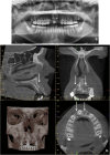
CBCT examination combined with 3D photography. (Courtesy of the manufacturer. The image is shown with the patient's permission)
Fig. 10
Fig. 10
Virtual implant treatment planning with CBCT data: software can help implant treatment planning through simulation and 3D reformation. (Courtesy Jari Mauno.)
Fig. 11
Fig. 11
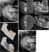
a Cropped PTG shows mesiocaudal dislocation of d. 47 with the root tips projecting into the lower cortex of the mandible. Distocranially to the crown of d. 47 a large complex odontoma is visible. b Based on the CBCT examination the mandibular canal could be located between the buccal and lingual roots (arrows); the root tips are in contact apically. c Rapid prototyping models based on the CBCT examination. d Postoperative cropped PTG: the roots of d. 47 were not removed in order to avoid nerve damage of the mandibular nerve bundle
Fig. 12
Fig. 12
a A patient with a unilateral cleft lip and palate has an alveolar defect on the left side (arrow). A PTG image taken before secondary alveolar bone grafting (SABG). Note also crowding in region d. 13, which can also be easily evaluated in the CBCT examination. b CBCT for the treatment planning before SABG allowing evaluation of the bone defect with oronasal fistula (arrows)
Fig. 13
Fig. 13
A large KCOT has been operated on in the left maxillary sinus. CBCT examination revealed a recurrence distally to d. 27, which was histopathologically proven (horizontal arrows). Note also the postoperative defect anteriorly and mucosal swelling in the maxillary sinus (vertical arrows)
Fig. 14
Fig. 14
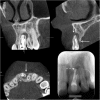
A deformed d. 21 was treated endodontically and resected several years ago. Clinically a fistula in the region d. 21 was found and fistulography with periapical radiography was done showing an apical periodontitis lesion (lower right). For the treatment planning a CBCT examination was done showing a large lesion indicating apical periodontitis with perforation of the labial cortex. Palatinally a vertical bone pocket is evident. Based on the CBCT findings extraction of the d. 21 was planned followed by implant treatment
Fig. 15
Fig. 15
The patient had been in a motorcycle accident. (a) Dd. 12, 21 and 22 have been exarticulated and their root sockets are visible in the PTG (horizontal arrows). Alveolar fracture (dashed arrow) is not so easily visible in PTG in comparison to the CBCT examination where the alveolar fracture with dislocation is evident (vertical arrows) (b). (Courtesy Tapio Tammisalo.)

References

    1. Boeddinghaus R, Whyte A. Current concepts in maxillofacial imaging. Eur J Radiol. 2008;66:396–418. doi: 10.1016/j.ejrad.2007.11.019. - DOI - PubMed
    1. Mozzo P, Procacci C, Tacconi A, Martini PT, Andreis IA. A new volumetric CT machine for dental imaging based on the cone-beam technique: preliminary results. Eur Radiol. 1998;8:1558–1564. doi: 10.1007/s003300050586. - DOI - PubMed
    1. Arai Y, Tammisalo E, Iwai K, Hashimoto K, Shinoda K. Development of a compact computed tomographic apparatus for dental use. Dentomaxillofac Radiol. 1999;28:245–248. doi: 10.1038/sj.dmfr.4600448. - DOI - PubMed
    1. Carter L, Farman AG, Geist J, Scarfe WC, Angelopoulos C, Nair MK, et al. American Academy of Oral and Maxillofacial Radiology executive opinion statement on performing and interpreting diagnostic cone beam computed tomography. Oral Surg Oral Med Oral Pathol Oral Radiol Endod. 2008 - PubMed
    1. Horner K, Islam M, Flygare L, Tsiklakis K, Whaites E. Basic principles for use of dental cone beam computed tomography: consensus guidelines of the European Academy of Dental and Maxillofacial Radiology. Dentomaxillofac Radiol. 2009 - PubMed