. 2015 May 15:574:108-19.
doi: 10.1016/j.abb.2014.12.025. Epub 2015 Jan 7.

Independent evolution of four heme peroxidase superfamilies

Affiliations

Independent evolution of four heme peroxidase superfamilies

Marcel Zámocký et al. Arch Biochem Biophys. .

Abstract

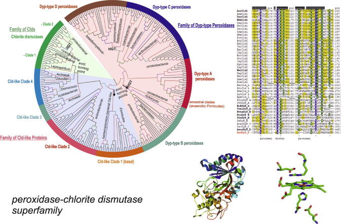
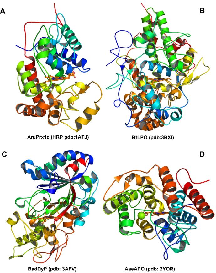
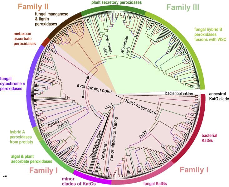
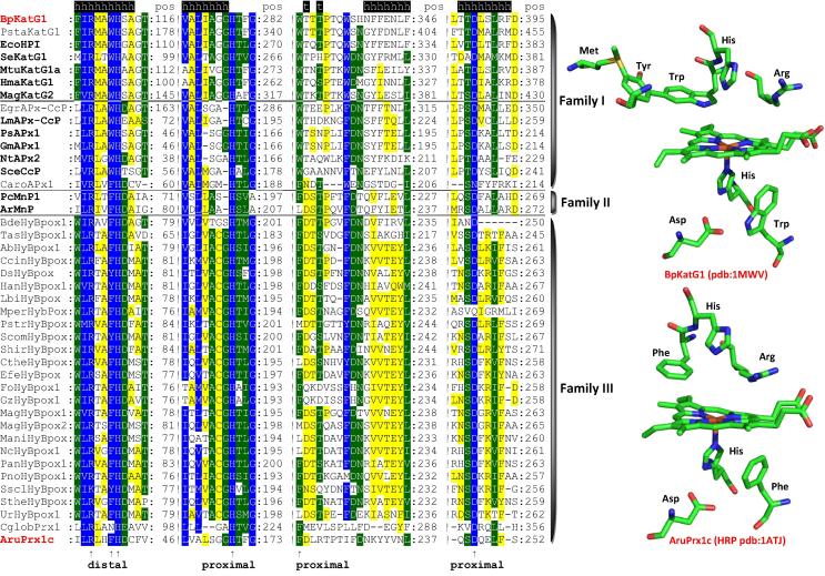
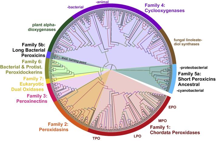
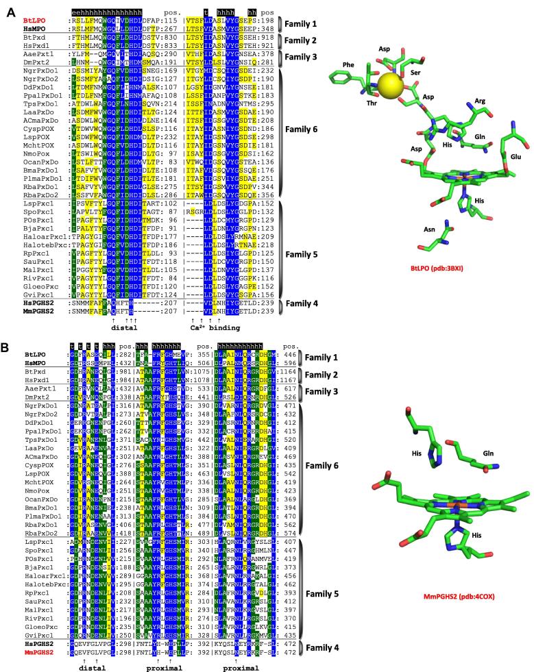
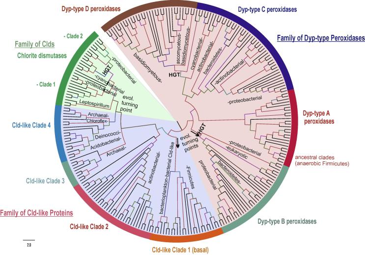
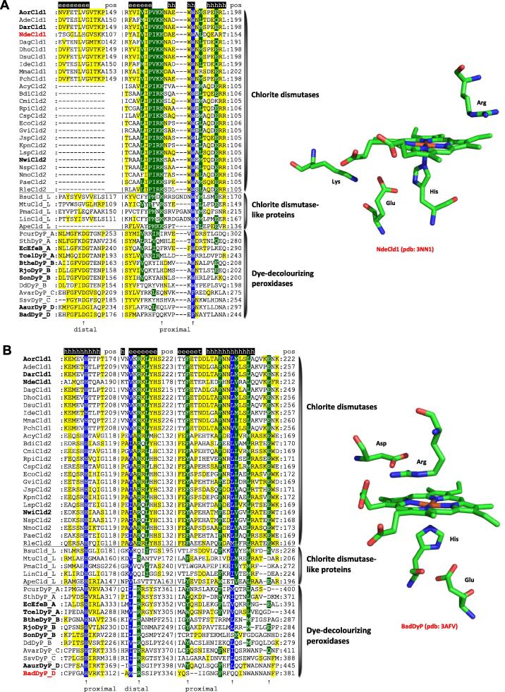
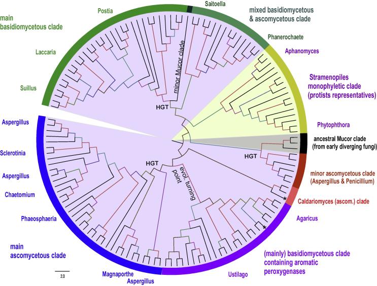
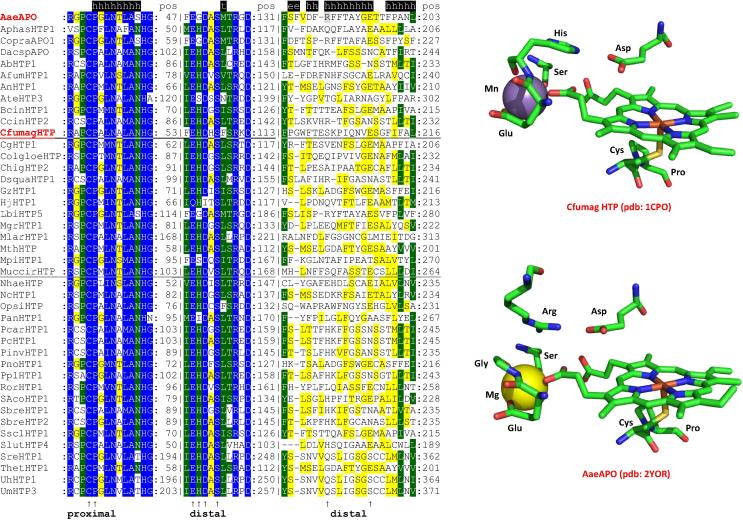
Four heme peroxidase superfamilies (peroxidase-catalase, peroxidase-cyclooxygenase, peroxidase-chlorite dismutase and peroxidase-peroxygenase superfamily) arose independently during evolution, which differ in overall fold, active site architecture and enzymatic activities. The redox cofactor is heme b or posttranslationally modified heme that is ligated by either histidine or cysteine. Heme peroxidases are found in all kingdoms of life and typically catalyze the one- and two-electron oxidation of a myriad of organic and inorganic substrates. In addition to this peroxidatic activity distinct (sub)families show pronounced catalase, cyclooxygenase, chlorite dismutase or peroxygenase activities. Here we describe the phylogeny of these four superfamilies and present the most important sequence signatures and active site architectures. The classification of families is described as well as important turning points in evolution. We show that at least three heme peroxidase superfamilies have ancient prokaryotic roots with several alternative ways of divergent evolution. In later evolutionary steps, they almost always produced highly evolved and specialized clades of peroxidases in eukaryotic kingdoms with a significant portion of such genes involved in coding various fusion proteins with novel physiological functions.

Keywords: Heme peroxidase; Peroxidase–catalase superfamily; Peroxidase–chlorite dismutase superfamily; Peroxidase–cyclooxygenase superfamily; Peroxidase–peroxygenase superfamily.


Figures

None
Graphical abstract
Fig. 1
Fig. 1
Structural fold typical for the peroxidase domains of each presented heme peroxidase superfamily. Representative structures with prosthetic heme group are shown for a typical member of (A) the peroxidase–catalase superfamily, (B) the peroxidase–cyclooxygenase superfamily, (C) the peroxidase–chlorite dismutase superfamily and (D) the peroxidase–peroxygenase superfamily. Abbreviations of presented peroxidases correspond with PeroxiBase and they are also explained in Supplem. Table 1.
Fig. 2
Fig. 2
Reconstructed phylogenetic tree of 500 members of the peroxidase–catalase superfamily. The evolutionary history was inferred by using the maximum likelihood method based on the Whelan & Goldman model implemented in MEGA 5 software . Bootstrap values are presented as color branches based on the obtained ML output: red > 90, violet > 70, blue > 50 and green > 30. All three main families (based on previously defined structural classes I, II and III known from [5]) are highlighted on the perimeter. Important subfamilies including the two hybrid-types are also highlighted.
Fig. 3
Fig. 3
Typical sequence patterns including distal and proximal triad found in all representatives of the peroxidase–catalase superfamily. In addition, the active site architectures of the Family I member, catalase–peroxidase from Burkholderia pseudomallei (BpKatG1) and the Family III protein horseradish peroxidase (AruPrx1c) is depicted. Note that BpKatG1 shows the unique KatG-typical distal side adduct that includes the distal tryptophan. Abbreviations of sequence names are explained in Supplem. Table 1 and they correspond with the nomenclature of PeroxiBase (http://peroxibase.toulouse.inra.fr). Color scheme: blue highest sequence similarity (93% conservation), green moderate similarity (75%), yellow low similarity (33%). Secondary structure elements for BpKatG1 structure are given above the alignment.
Fig. 4
Fig. 4
Reconstructed phylogenetic tree of 400 members of the peroxidase–cyclooxygenase superfamily. The evolutionary history was inferred by using the maximum likelihood method based on the Whelan & Goldman model implemented in MEGA 5 software . Bootstrap values are presented as a color branches based on the ML output: red > 90, violet > 70, blue > 50 and green > 30. All seven (numbered) families known from are highlighted on the perimeter together with important subfamilies.
Fig. 5
Fig. 5
Typical sequence pattern including distal and proximal residues found in representatives of the peroxidase–cyclooxygenase superfamily (except dual oxidases that were shown to contain a mutated peroxidase domain without peroxidase activity . (A) Distal heme side and calcium binding motif, (B) further distal motif and proximal heme side. Color scheme: blue highest similarity (93% conservation), green moderate similarity (70%), yellow low similarity (33%). In addition the active site architectures of bovine lactoperoxidase and mouse prostaglandin H synthase 2 are depicted. Abbreviations of sequence names are explained in Supplem. Table 1 and they correspond with the nomenclature of PeroxiBase (http://peroxibase.toulouse.inra.fr). Secondary structure elements for BtLPO structure are given above the alignment.
Fig. 6
Fig. 6
Reconstructed phylogenetic tree of 250 members of the peroxidase–chlorite dismutase superfamily. The evolutionary history was inferred by using the maximum likelihood method based on the Whelan & Goldman model implemented in MEGA 5 software . Bootstrap values are presented as color branches based on the ML output: red > 90, violet > 70, blue > 50 and green > 30. Three main families [chlorite dismutases (Clds), dye-decolorizing peroxidases (DyPs) and Cld-like proteins] known from are highlighted on the perimeter. Important subfamilies (in some cases clades) are also shown.
Fig. 7
Fig. 7
Typical sequence pattern including distal and proximal residues found in representatives of the peroxidase–chlorite dismutase superfamily. Chlorite dismutases (Clds), dye-decolorizing peroxidases (DyPs) and Cld-like proteins. Color scheme: blue highest similarity (93% conservation), green moderate similarity (67%), yellow low similarity (30%). In addition the active site architectures of chlorite dismutase from Nitrospira defluvii (Clade 1 Cld) and of DyP from Bjerkandera adusta (Dyp-type D) are depicted. Abbreviations of sequence names are explained in Supplem. Table 1 and for peroxidase representatives correspond with the nomenclature of PeroxiBase (http://peroxibase.toulouse.inra.fr). Secondary structure elements for NdeCld1 structure are given above the alignment.
Fig. 8
Fig. 8
Reconstructed phylogenetic tree of 136 members of the peroxidase–peroxygenase superfamily. The evolutionary history was inferred by using the maximum likelihood method based on the Whelan & Goldman model implemented in MEGA 5 software . Bootstrap values are presented as color branches based on the ML output: red > 90, violet > 70, blue > 50 and green > 30. All main subfamilies (currently defined only as clades) are highlighted on the perimeter.
Fig. 9
Fig. 9
Typical sequence pattern including distal and proximal residues found in representatives of the peroxidase–peroxygenase superfamily. Color scheme: blue highest similarity (93% conservation), green moderate similarity (67%), yellow low similarity (33%). In addition the active site architectures of enzymes from Caldariomyces fumago and Agrocybe aegerita are depicted. Note that both proteins have a cation-binding site in the heme periphery. Abbreviations of sequence names are explained in Supplem. Table 1 and they correspond with the nomenclature of PeroxiBase (http://peroxibase.toulouse.inra.fr). Secondary structure elements for AaeAPO structure are given above the alignment.

References

    1. Sammut S.J., Finn R.D., Bateman A. Brief. Bioinf. 2008;9:210–219. - PubMed
    1. Chothia C., Cough J. Biochem. J. 2009;419:15–28. - PubMed
    1. Caetano-Anolles G., Wang M., Caetano-Anolles D., Mittenthal J.E. Biochem. J. 2009;417:621–637. - PubMed
    1. Zamocky M., Jakopitsch C., Furtmüller P.G., Dunand C., Obinger C. Proteins. 2008;72:589–605. - PubMed
    1. Welinder K.G. Curr. Opin. Struct. Biol. 1992;2:388–393.

Publication types