Case Reports
. 1989;9(6):633-54.
doi: 10.3109/15513818909022372.

Multivisceral intestinal transplantation: surgical pathology

Affiliations
Case Reports

Multivisceral intestinal transplantation: surgical pathology

R Jaffe et al. Pediatr Pathol. 1989.

Abstract

We report the diagnostic surgical pathology of two children who underwent multivisceral abdominal transplantation and survived for 1 month and 6 months. There is little relevant literature, and diagnostic criteria for the various clinical possibilities are not established; this is made more complicated by the simultaneous occurrence of more than one process. We based our interpretations on conventional histology, augmented with immunohistology, including HLA staining that distinguished graft from host cells in situ. In some instances functional analysis of T cells propagated from the same biopsies was available and was used to corroborate morphological interpretations. A wide spectrum of changes was encountered. Graft-versus-host disease, a prime concern before surgery, was not seen. Rejection was severe in 1 patient, not present in the other, and both had evidence of lymphoproliferative disease, which was related to Epstein-Barr virus. Bacterial translocation through the gut wall was also a feature in both children. This paper documents and illustrates the various diagnostic possibilities.


Figures
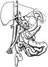
FIGURE 1
FIGURE 1
The recipient operation of multivisceral transplantation. Note that the venous outflow of the graft was into a cloaca of the left and middle hepatic veins, leaving the recipient vena cava intact. (A: donor aorta; HA: hepatic artery; SA: splenic artery; LGA: left gastric artery; SMA: superior mesentric artery; IMA: inferior mesenteric artery; GDA: gastroduodenal artery.)
FIGURE 2
FIGURE 2
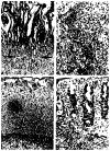
Intestinal sections, patient 1. (a) Donor colon; biopsy at 6 months. The epithelium is intact; there is no cellular infiltrate. × 240. (b) Donor jejunum; postmortem sample. Hyperplastic villi have intact epithelium. ×120. (c)Donor midileum; postmortem sample; dystrophic changes. The mucosa has partial villus atrophy, and the submucosa is fibrotic; no cellular infiltrate is present. × 120. (d) Donor terminal ileum. The surface is lined by a single layer of regenerating columnar cells. The submucosa and muscularis arc scarred. × 120.
FIGURE 3
FIGURE 3
Intestinal and liver sections, patient 2, day 24. (a) Donor stomach; rejection. The gastric mucosa is tall, but the deep glands are being destroyed by a cellular infiltrate (arrows)× 120. (b) Donor liver; rejection. The portal area is expanded by a mixed cellular infiltrate rich in lymphocytes and eosinophils. The hepatic lobule is relatively spared. × 280. (c)Donor, terminal ileum; rejection. The mucosa is devoid of epithelium. A residual lymphoid follicle is recognizable. The lamina propria is populated by lymphoid cells. × 160. (d) Donor colon; rejection. Damaged and regenerating glands and surface epithelium are present, and an infiltrate separates the glands. ×160.
FIGURE 4
FIGURE 4
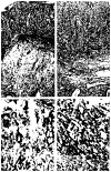
Donor colon, frozen sections, patient 2, day 27. (a) Donor-specific antibody Bw6 stains epithelial and endothelial cells but not the cellular infiltrate. (b) Recipient specific antibody HLA 25/32 give a mirror image to (a); the infiltrating cells in the lamina propria are of recipient phenotype (c) HLA-DR staining reveals intense expression on epithelial as well as infiltrating cells. Surprisingly, some glands (arrow) are unstained. (d) Many of the infiltrating lymphoid cells bear CD25 (IL-2) receptors × 220.
FIGURE 5
FIGURE 5
Donor mesenteric node, patient 2, day 24. (a) Marked paracortical expansion compresses the small follicles against the capsule. Giemsa, ×160. (b) The exuberant proliferation of interdigitating dendritic cells is revealed in the paracortex by S-100 staining. × 440.
FIGURE 6
FIGURE 6
Lymphoproliferative disease, patient I. (a) Biopsy of the liver at 91 days reveals a fairly uniform population of cells that obscure all portal landmarks and spills over into the lobule. Giemsa, × 110. (b) The lymphoproliferative reaction was of recipient phenotype. Recipient specific antibody (R) HLA-A3 stains most cells, whereas the donor specific antibody (D) Bw4 leaves the cells unstained. × 200. (c) Although immunoglobulin cells predominate, T cells, as demonstrated by UCH-l, are well represented in the lesion. × 200. (d) The calcific rim is seen around the ghost cells of what appears to be infarcted lymphoproliferative disease at day 137. × 180.
FIGURE 7
FIGURE 7
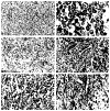
Liver, postmortem, patient I. (a) A large juxtahilar mass consists in part of lymphoproliferative disease and infarction. (b) The presumably lymphoid proliferation is perivascular, angiocentric, and angioinvasive. × 150. (c) The wall of a hepatic artery is infiltrated and destroyed by the atypical cellular proliferation. × 350. (d) The cells did not react with B cell, T cell, or monocyte markers. S100 reveals a nerve, but the cells are unreactive. They are morphologically different from the plasmacytoid elements that were still recognizable elsewhere. × 660.
FIGURE 8
FIGURE 8
Lymphoproliferative disease, patient 2. (a) Donor ileum, day 24; rejection. Severe cellular rejection is represented. Fragments of residual epithelium are seen, and a mixed cellular infiltrate is presented in the lamina propria, × 160. (b) Donor ileum, day 34; lymphoproliferative disease. The infiltrate is more exuberant, more monotonous, the cells are larger, and the infiltrate obscures anatomic boundaries such as the muscularis mucosae, × 160. (c) Ileum, day 34. Frozen section stained with donor specific antibody Bw6 stains endothelial cells and epithelial remnants, × 300. (d) An adjacent section stained with recipient-specific antibody HLA 25/32 highlights the infiltrating lymphoid cells. The pattern does not distinguish rejection (see Fig. 4) from the lymphoproliferative process, ×300.
FIGURE 9
FIGURE 9
Lymphoproliferative disease; donor mesenteric lymph node, day 34, patient 2 (compare with day 24, Fig. 5). (a) Whereas the lymph node previously demonstrated an expanded paracortex, the lymph node architecture is now effaced by a diffuse lymphoid proliferation. × 120. (b) The proliferation is polymorphous; lymphocytes, plasma cells, plasmacytoid cells, and immunoblasts are represented. Giemsa, × 660. (c) Donor lymph node, day 34 (see Fig. 9). Most of the immunoglobulin-containing cells stain for IgM. Kappa and lambda chains were equally represented. Immunoperoxidase, × 160. (d) The lymphoproliferative process was not confined to donor organs, being seen here in the host kidney. × 280. (e) Recipient-specific antibody stains almost all infiltrating cells but not endothelium or sinus lining cells. Frozen section, immunoperoxidase. × 300. (f) Donor-specific antibody Bw6 stains sinus lining cells, endothelial cells, and occasional large (possibly dendritic) cells. × 300.
FIGURE 10
FIGURE 10
(a) Donor mesenteric lymph node at day 17. 35S label for EBV DNA is seen in a lymphoid follicle (arrow). (The large black object at center is artefact.) × 200. (b) Donor mesentric lymph node at day 34. There is now diffuse labeling for EBV DNA of many cells throughout the node. × 200.

References

    1. Starzl TE, Rowe MI, Todo S, et al. Transplantation of multiple abdominal viscera. JAMA. 1989;261:1449–57. - PMC - PubMed
    1. Williams JS, Sankary HN, Foster PF, Lowe J, Goldman GM. Splanchnic transplantation: An approach to the infant dependent on parenteral nutrition who develops irreversible liver disease. JAMA. 1989;261:1458–62. - PubMed
    1. Goulet OJ, Révillon Y, Cerf-Bensussan N, et al. Small intestinal transplantation in a child using cyclosporine. Transpl Proc. 1988;20:288–96. - PubMed
    1. Cutz E, Rhoads JM, Drumm B, Sherman PM, Durie PR, Forstner GG. Microvillus inclusion disease: An inherited defect of brush border assembly and differentiation. N Engl J Med. 1989;320:646–51. - PubMed
    1. Todo S, Nery J, Yanaga K, Podesta L, Gordon RD, Starzl TE. Extended preservation of human liver grafts with UW solution. JAMA. 1989;261:711–4. - PMC - PubMed

Publication types

MeSH terms

Substances