. 2015 Mar;167(3):738-52.
doi: 10.1104/pp.114.254938. Epub 2015 Jan 9.

Importin-α-mediated nucleolar localization of potato mop-top virus TRIPLE GENE BLOCK1 (TGB1) protein facilitates virus systemic movement, whereas TGB1 self-interaction is required for cell-to-cell movement in Nicotiana benthamiana

Affiliations

Importin-α-mediated nucleolar localization of potato mop-top virus TRIPLE GENE BLOCK1 (TGB1) protein facilitates virus systemic movement, whereas TGB1 self-interaction is required for cell-to-cell movement in Nicotiana benthamiana

Nina I Lukhovitskaya et al. Plant Physiol. 2015 Mar.

Abstract

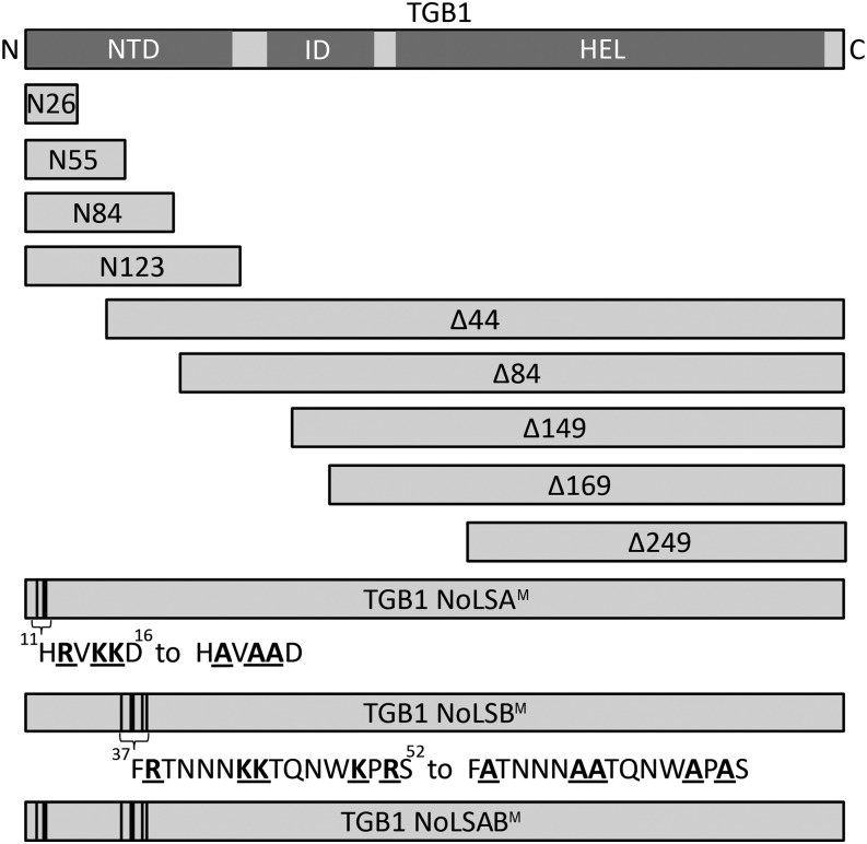
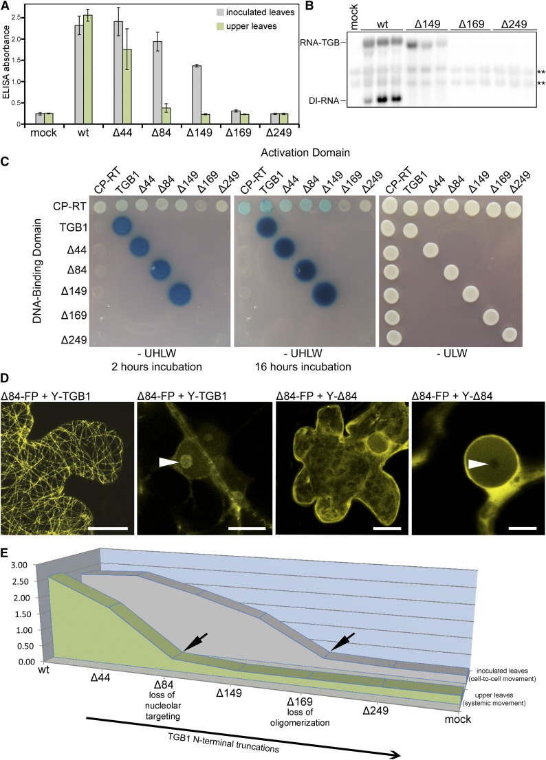
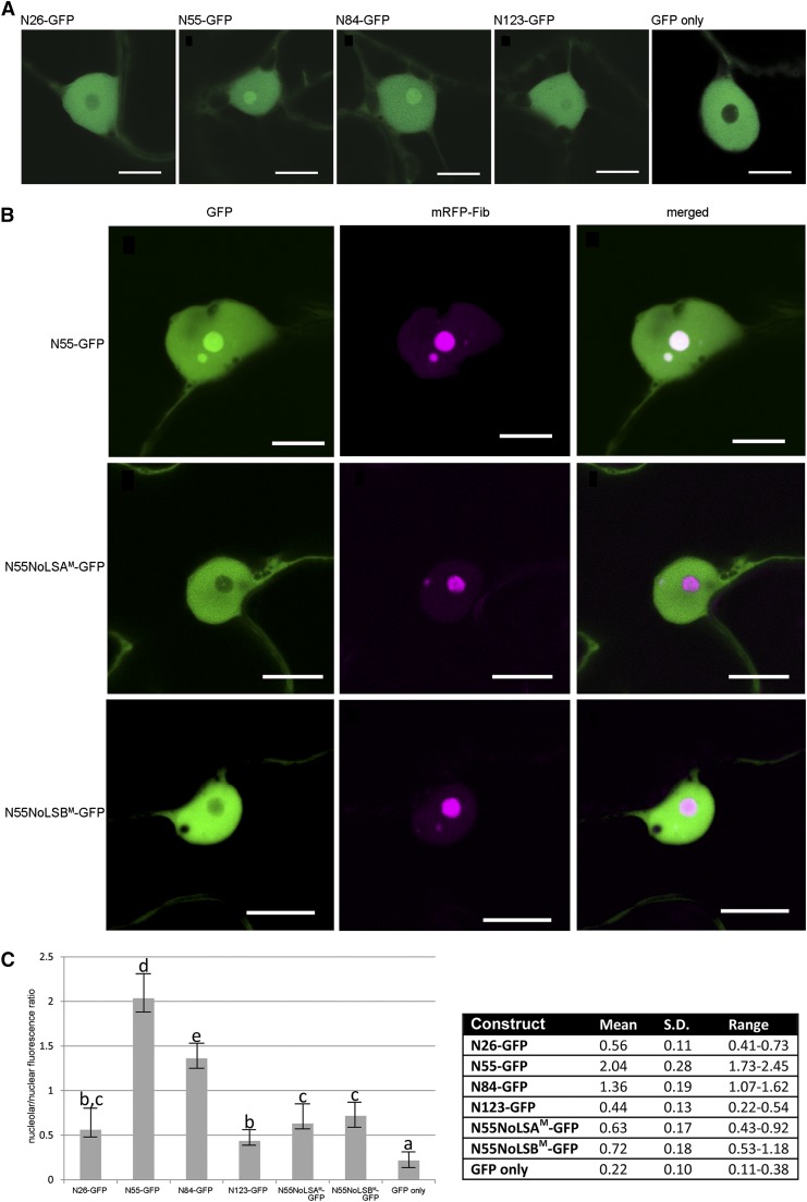
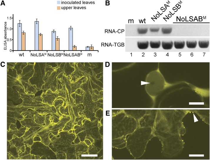
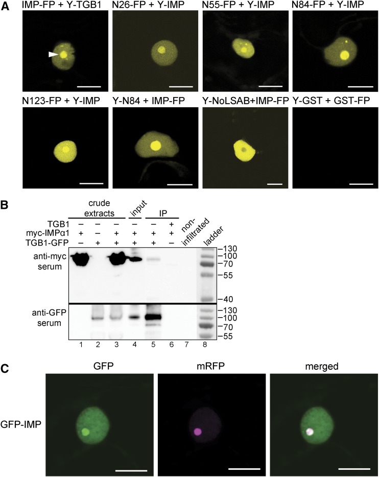
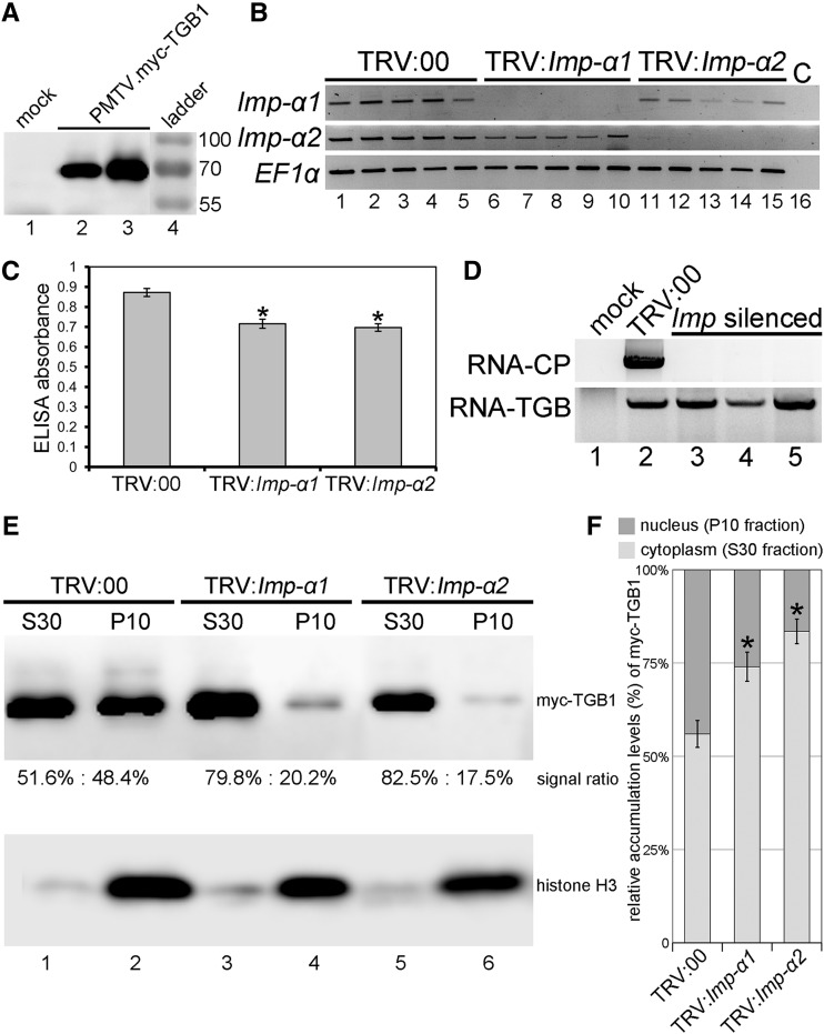
Recently, it has become evident that nucleolar passage of movement proteins occurs commonly in a number of plant RNA viruses that replicate in the cytoplasm. Systemic movement of Potato mop-top virus (PMTV) involves two viral transport forms represented by a complex of viral RNA and TRIPLE GENE BLOCK1 (TGB1) movement protein and by polar virions that contain the minor coat protein and TGB1 attached to one extremity. The integrity of polar virions ensures the efficient movement of RNA-CP, which encodes the virus coat protein. Here, we report the involvement of nuclear transport receptors belonging to the importin-α family in nucleolar accumulation of the PMTV TGB1 protein and, subsequently, in the systemic movement of the virus. Virus-induced gene silencing of two importin-α paralogs in Nicotiana benthamiana resulted in significant reduction of TGB1 accumulation in the nucleus, decreasing the accumulation of the virus progeny in upper leaves and the loss of systemic movement of RNA-CP. PMTV TGB1 interacted with importin-α in N. benthamiana, which was detected by bimolecular fluorescence complementation in the nucleoplasm and nucleolus. The interaction was mediated by two nucleolar localization signals identified by bioinformatics and mutagenesis in the TGB1 amino-terminal domain. Our results showed that while TGB1 self-interaction is needed for cell-to-cell movement, importin-α-mediated nucleolar targeting of TGB1 is an essential step in establishing the efficient systemic infection of the entire plant. These results enabled the identification of two separate domains in TGB1: an internal domain required for TGB1 self-interaction and cell-to-cell movement and the amino-terminal domain required for importin-α interaction in plants, nucleolar targeting, and long-distance movement.


Figures

Figure 1.
Figure 1.
Schematic representation of the TGB1 constructs used in this study. Schematics are shown for full-length TGB1, N-terminal truncations (Δ), N-terminal fragments (N), and amino acid substitutions in the NoLS of PMTV TGB1 analyzed in this article. At top is the domain structure of the PMTV TGB1 protein. The N-terminal fragments and N-terminal truncations are shown to scale. Superscript numbers refer to amino acid residue positions within the sequence of the TGB1 protein. ID, Internal domain; HEL, helicase domain.
Figure 2.
Figure 2.
Effects of N-terminal truncations in the PMTV TGB1 protein on viral RNA cell-to-cell and long-distance movement and self-interaction of TGB1. A, Detection of PMTV by ELISA as presented by absorbance values at 405 nm. Plant extracts were prepared from inoculated and upper leaves 14 dpi. The average absorbance of a healthy plant extract was equal to 0.25. The data are from two independent experiments (n = 10); error bars denote sd. B, RNA gel-blot analyses of the accumulation of wild-type PMTV (wt) and the PMTV TGB1 mutants in inoculated leaves of N. benthamiana. Total RNA was isolated 14 dpi. RNA-TGB was detected using 32P-labled 5′ untranslated region antisense RNA probe. Asterisks indicate nonspecific binding of the probe. C, Yeast two-hybrid assay for studying self-interactions and CP-RT interactions of TGB1 and its N terminally truncated forms. Wild-type TGB1 or the TGB1 N-terminal truncated sequences were fused to the LexA-DNA-binding domain or the herpes simplex virus protein16 (VP16) transcription activation domain. Transformants were selected for protein interactions on medium lacking uracil, His, Leu, and Trp (–UHLW), a triple dropout medium. Medium lacking uracil, Leu, and Trp (–ULW), a double dropout medium, selected for the input plasmids only and shows growth of the colonies carrying both plasmids as a positive control. As reported previously (Wright et al., 2010), interaction between TGB1 variants and CP-RT could only be detected when CP-RT was fused to the DNA-binding domain and TGB1 to the activation domain (horizontal row at top) but not in the reverse orientation (vertical row at left), possibly due to steric hindrance. β-Galactosidase activity was assessed by agarose overlay assays at two time points (at 2 and 16 h of incubation at 28°C). D, Interaction of full-length TGB1 and Δ84, and self-interaction of Δ84, detected by BiFC. Interacting TGB1 and Δ84 decorate MTs and localize to the nucleolus (arrowhead), whereas self-interacting Δ84 does not localize to MTs, resides mostly in the cytoplasm, and is excluded from the nucleolus (arrowhead). Bars = 10 μm (first three images from left) and 5 μm (fourth image). E, Three-dimensional chart showing the correlation between TGB1 oligomerization, nucleolar targeting, and virus accumulation in inoculated (due to cell-to-cell movement) and upper (due to systemic movement) leaves.
Figure 3.
Figure 3.
Nuclear localization of TGB1 N-terminal fragments fused to GFP. A, Accumulation of TGB1 N-terminal fragments (26, 55, 84, and 123 amino acid residues) fused to GFP in the nucleus and their partial exclusion from the nucleolus (N26-GFP and N123-GFP). N55-GFP and N84-GFP accumulate in the nucleoplasm and are enriched within the nucleolus. Bars = 5 μm. B, TGB1 N55-GFP fusion and its two derivatives with amino acids substitutions in the NoLSA and NoLSB motifs (N55NoLSAM-GFP and N55NoLSBM-GFP, respectively). GFP is shown in green, and the nucleolar marker, mRFP-Fib, is shown in magenta. Bars = 5 μm. C, Quantification of the nucleolar-nuclear fluorescence ratio for the four N-terminal fragments as well as N55NoLSAM and N55NoLSBM. Error bars represent sd (n = 10), and letters indicate groups of constructs that differ significantly from each other (one way-ANOVA and Dunnett’s T3 test; P < 0.001). The table at right shows mean, sd, and range of fluorescence ratios.
Figure 4.
Figure 4.
Effects of the Ala substitutions of the Arg and Lys residues in NoLSA, NoLSB, and both NoLS motifs on TGB1 localization and virus accumulation in inoculated and upper leaves. A, Accumulation of PMTV measured by ELISA as indicated by absorbance values at 405 nm. Plant extracts were prepared from inoculated and upper leaves 14 dpi. The average absorbance of a healthy plant extract (m, mock) was equal to 0.17. ELISA experiments were conducted twice (n = 6). Error bars denote sd. wt, Wild type. B, RT-PCR on RNA of N. benthamiana upper leaves to detect the accumulation of viral RNA-CP and RNA-TGB. The identity of each virus inoculum is indicated above the gels (lanes 5–7 are different plants inoculated with PMTV-NoLSABM). Thirty cycles were used for amplification. The experiment was repeated twice with similar results. C, Multicellular lesion of PMTV.YFP-TGB1NoLSABM 14 dpi showing cell-to-cell movement of the mutant virus. Bar = 40 µm. D, Localization of YFP-TGB1NoLSABM to the nucleolus upon expression from the viral genome. Bar = 5 µm. E, Single confocal section illustrating YFP-TGB1NoLSABM located in punctate structures (arrowhead) in the cell wall, presumably plasmodesmata. Bar = 5 µm.
Figure 5.
Figure 5.
Importin-α interacts with the PMTV TGB1 protein. Interactions were assayed by BiFC and co-IP. A, TGB1 fused to the N-terminal half of YFP and NbImp-α1 fused to the C-terminal half of YFP transiently coexpressed in epidermal cells of N. benthamiana show reconstituted fluorescence of YFP in the nucleoplasm and accumulation of fluorescence at the nucleolus. TGB1 N-terminal fragments (26, 55, 84, and 123 residues) fused to the C-terminal half of YFP and NbImp-α1 fused to the N-terminal half of YFP (Y-IMP) show reconstituted YFP fluorescence in the nucleoplasm and accumulation at the nucleolus. Cells coexpressing TGB1 altered in both NoLSs (NoLSABM) fused to the N-terminal half of YFP and NbImp-α1 fused to the C-terminal half of YFP show reconstituted YFP fluorescence in the nucleoplasm and partial exclusion from the nucleolus. The BiFC control with GST fused to the N-terminal and C-terminal halves of YFP shows no reconstituted fluorescence. For all tested BiFC combinations, see Table III. B, Co-IP of extracts from N. benthamiana leaves coinfiltrated with TGB1-GFP and myc-IMPα1, or TGB1 and myc-IMPα1, using anti-GFP microbeads, followed by immunoblot analysis with anti-myc and anti-GFP antibodies (lanes 4–6). The coexpression of nonfused TGB1 and myc-IMPα1 was used as a control in the co-IP experiment (expression controls for the proteins; lanes 1–3). The experiments were conducted twice. C, GFP-IMPα1 (green) coexpression with mRFP-Fib (magenta). Bars = 5 μm.
Figure 6.
Figure 6.
Effects of the knockdown of the expression of two importin-α homologs on PMTV accumulation in upper noninoculated leaves. A, Immunoblot analysis of c-myc epitope-tagged TGB1 expressed from the virus. At 14 dpi, samples from upper noninoculated leaves were analyzed by immunoblot using anti-myc antibody. Positions of protein standards (in kD) are marked at right. B, RT-PCR of RNA from 4-week-old N. benthamiana plants inoculated with TRV:Imp-α1 or TRV:Imp-α2 silencing constructs or with an empty VIGS vector (TRV:00). Primers detect Importin-α1, Importin-α2, or EF-1α transcripts. Each lane corresponds to a different plant. C, Levels of PMTV accumulation detected by ELISA as indicated by absorbance values at 405 nm. Plant extracts were prepared from upper leaves 14 dpi. The average absorbance of a healthy plant extract was equal to 0.14. Asterisks indicate that the absorbance values were significantly different (P < 0.05; Student’s t test) from those of nonsilenced control plants (TRV:00). Experiments were conducted twice (n = 10). D, RT-PCR analysis of RNA from Importin-α1-silenced plants (lanes 3 and 4) and Importin-α2-silenced plants (lane 5), which were completely negative for the presence of PMTV CP antigen in ELISA, using primers to detect PMTV RNA-CP or PMTV RNA-TGB. RNA samples from the upper leaves of a mock-inoculated plant or from the upper leaves of a plant inoculated with empty VIGS vector (TRV:00) served as controls. E, Presence of c-myc-tagged TGB1 in the nuclear P10 and cytoplasmic S30 fractions. The fractions were analyzed by immunoblotting using antisera to the c-myc-tag (top) and to histone H3 as a nuclear marker (bottom). The relative extent of myc-TGB1 signal in the nuclear and cytoplasmic fractions is shown below the top gel. F, Relative myc-TGB1 accumulation levels were measured on immunoblots in two independent experiments (n = 5). Additional images used for quantification are shown in Supplemental Figure S2C. Asterisks indicate statistically significant differences compared with the control group, TRV:00 (P < 0.01; Student’s t test).

Similar articles

Cited by

References

    1. Arif M, Torrance L, Reavy B (1995) Acquisition and transmission of potato mop-top furovirus by a culture of Spongospora subterranea f.sp subterranea derived from a single cystosorus. Ann Appl Biol 126: 493–503
    1. Barneche F, Steinmetz F, Echeverría M (2000) Fibrillarin genes encode both a conserved nucleolar protein and a novel small nucleolar RNA involved in ribosomal RNA methylation in Arabidopsis thaliana. J Biol Chem 275: 27212–27220 - PubMed
    1. Chiba S, Hleibieh K, Delbianco A, Klein E, Ratti C, Ziegler-Graff V, Bouzoubaa S, Gilmer D (2013) The benyvirus RNA silencing suppressor is essential for long-distance movement, requires both zinc-finger and NoLS basic residues but not a nucleolar localization for its silencing-suppression activity. Mol Plant Microbe Interact 26: 168–181 - PubMed
    1. Crivelli G, Ciuffo M, Genre A, Masenga V, Turina M (2011) Reverse genetic analysis of Ourmiaviruses reveals the nucleolar localization of the coat protein in Nicotiana benthamiana and unusual requirements for virion formation. J Virol 85: 5091–5104 - PMC - PubMed
    1. Dosztányi Z, Csizmok V, Tompa P, Simon I (2005) IUPred: web server for the prediction of intrinsically unstructured regions of proteins based on estimated energy content. Bioinformatics 21: 3433–3434 - PubMed

Publication types

Substances

LinkOut - more resources