

Direct intracardiac injection of umbilical cord-derived stromal cells and umbilical cord blood-derived endothelial cells for the treatment of ischemic cardiomyopathy
- PMID: 25576340
- PMCID: PMC4935404
- DOI: 10.1177/1535370214565077
Direct intracardiac injection of umbilical cord-derived stromal cells and umbilical cord blood-derived endothelial cells for the treatment of ischemic cardiomyopathy
Abstract
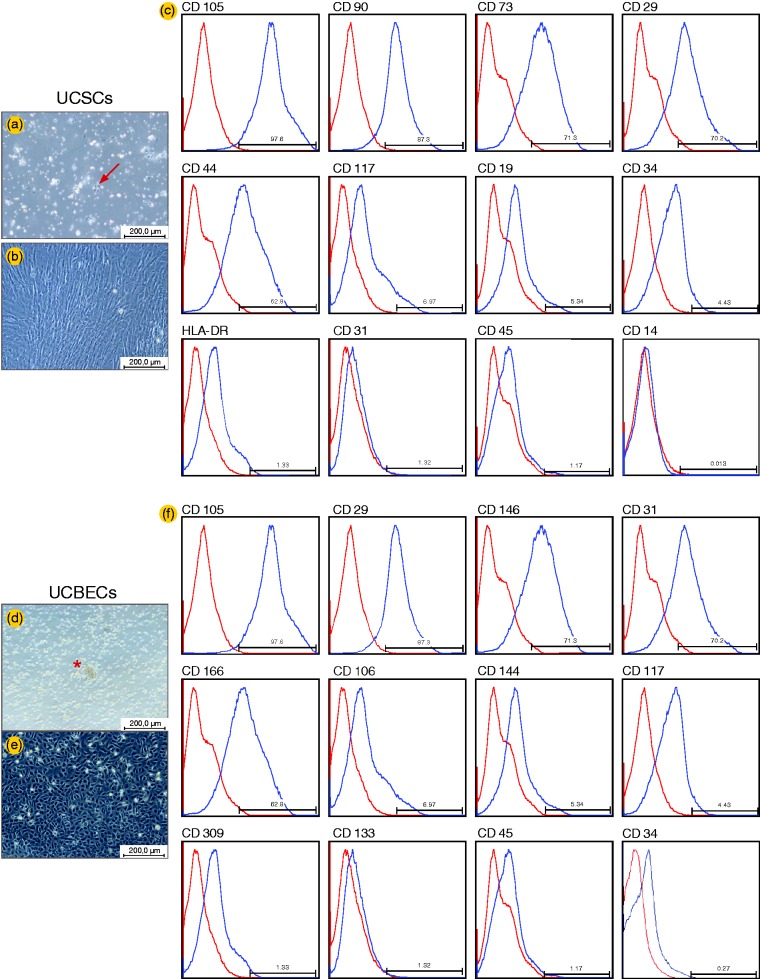
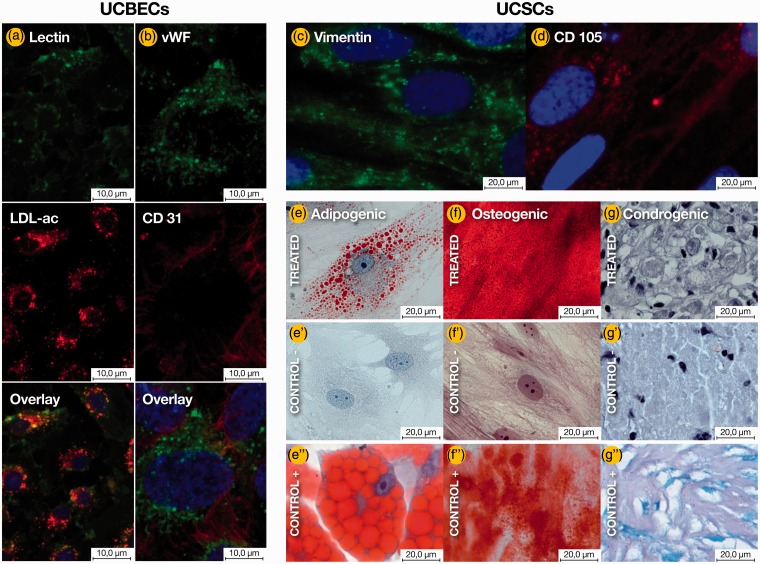
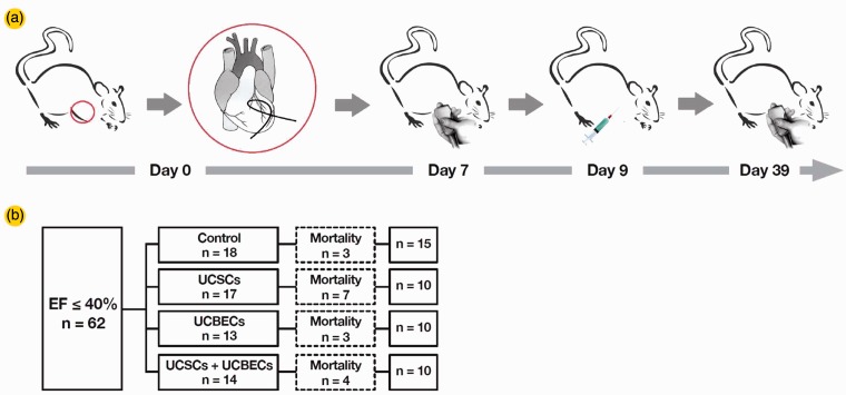
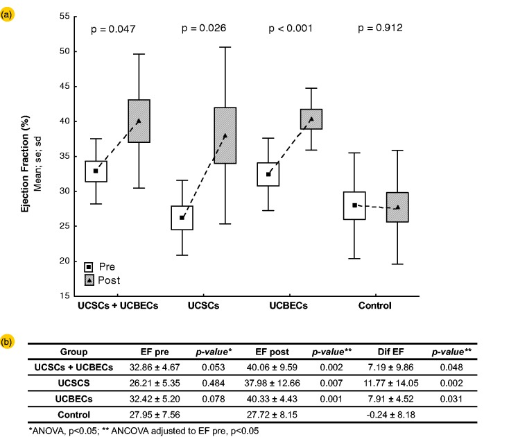
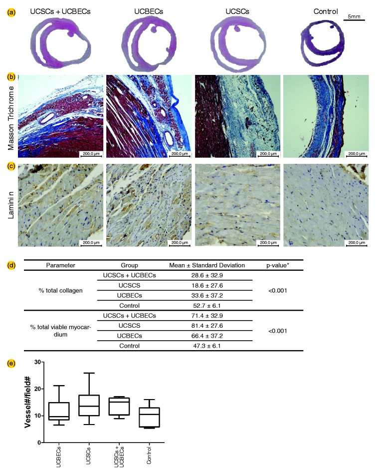
The development of new therapeutic strategies is necessary to reduce the worldwide social and economic impact of cardiovascular disease, which produces high rates of morbidity and mortality. A therapeutic option that has emerged in the last decade is cell therapy. The aim of this study was to compare the effect of transplanting human umbilical cord-derived stromal cells (UCSCs), human umbilical cord blood-derived endothelial cells (UCBECs) or a combination of these two cell types for the treatment of ischemic cardiomyopathy (IC) in a Wistar rat model. IC was induced by left coronary artery ligation, and baseline echocardiography was performed seven days later. Animals with a left ventricular ejection fraction (LVEF) of ≤40% were selected for the study. On the ninth day after IC was induced, the animals were randomized into the following experimental groups: UCSCs, UCBECs, UCSCs plus UCBECs, or vehicle (control). Thirty days after treatment, an echocardiographic analysis was performed, followed by euthanasia. The animals in all of the cell therapy groups, regardless of the cell type transplanted, had less collagen deposition in their heart tissue and demonstrated a significant improvement in myocardial function after IC. Furthermore, there was a trend of increasing numbers of blood vessels in the infarcted area. The median value of LVEF increased by 7.19% to 11.77%, whereas the control group decreased by 0.24%. These results suggest that UCSCs and UCBECs are promising cells for cellular cardiomyoplasty and can be an effective therapy for improving cardiac function following IC.
Keywords: Human umbilical cord-derived stromal cells; cell therapy; human umbilical cord blood-derived endothelial cells; ischemic cardiomyopathy.
© 2015 by the Society for Experimental Biology and Medicine.
Figures
Similar articles
-
Expanded human cord blood-derived endothelial progenitor cells salvage infarcted myocardium in rats with acute myocardial infarction.Clin Exp Pharmacol Physiol. 2010 May;37(5-6):551-6. doi: 10.1111/j.1440-1681.2010.05347.x. Clin Exp Pharmacol Physiol. 2010. PMID: 20529094
-
Transplanted human umbilical cord blood mononuclear cells improve left ventricular function through angiogenesis in myocardial infarction.Chin Med J (Engl). 2006 Sep 20;119(18):1499-506. Chin Med J (Engl). 2006. PMID: 16996002
-
Intraarterial transplantation of human umbilical cord blood mononuclear cells is more efficacious and safer compared with umbilical cord mesenchymal stromal cells in a rodent stroke model.Stem Cell Res Ther. 2014 Apr 1;5(2):45. doi: 10.1186/scrt434. Stem Cell Res Ther. 2014. PMID: 24690461 Free PMC article.
-
Umbilical cord blood-derived mesenchymal stem cells: new therapeutic weapons for idiopathic dilated cardiomyopathy?Int J Cardiol. 2014 Dec 20;177(3):809-18. doi: 10.1016/j.ijcard.2014.09.128. Epub 2014 Oct 2. Int J Cardiol. 2014. PMID: 25305679 Review.
-
Pluripotent possibilities: human umbilical cord blood cell treatment after neonatal brain injury.Pediatr Neurol. 2013 May;48(5):346-54. doi: 10.1016/j.pediatrneurol.2012.10.010. Pediatr Neurol. 2013. PMID: 23583051 Review.
Cited by
-
Treatment of Chronic Kidney Disease with Extracellular Vesicles from Mesenchymal Stem Cells and CD133+ Expanded Cells: A Comparative Preclinical Analysis.Int J Mol Sci. 2022 Feb 25;23(5):2521. doi: 10.3390/ijms23052521. Int J Mol Sci. 2022. PMID: 35269664 Free PMC article.
-
Systemic Infusion of Expanded CD133+ Cells and Expanded CD133+ Cell-Derived EVs for the Treatment of Ischemic Cardiomyopathy in a Rat Model of AMI.Stem Cells Int. 2019 Dec 1;2019:4802578. doi: 10.1155/2019/4802578. eCollection 2019. Stem Cells Int. 2019. PMID: 31885610 Free PMC article.
-
Forecasting mergers and acquisitions failure based on partial-sigmoid neural network and feature selection.PLoS One. 2021 Nov 17;16(11):e0259575. doi: 10.1371/journal.pone.0259575. eCollection 2021. PLoS One. 2021. PMID: 34788332 Free PMC article.
-
Expanded CD133+ Cells from Human Umbilical Cord Blood Improved Heart Function in Rats after Severe Myocardial Infarction.Stem Cells Int. 2018 Apr 11;2018:5412478. doi: 10.1155/2018/5412478. eCollection 2018. Stem Cells Int. 2018. PMID: 29760727 Free PMC article.
-
Mesenchymal stem cells in ischemic tissue regeneration.World J Stem Cells. 2023 Feb 26;15(2):16-30. doi: 10.4252/wjsc.v15.i2.16. World J Stem Cells. 2023. PMID: 36909782 Free PMC article. Review.
References
-
- Lopez AD, Mathers CD, Ezzati M, Jamison DT, Murray CJ. Global and regional burden of disease and risk factors, 2001: systematic analysis of population health data. Lancet 2006; 367: 1747–57. - PubMed
-
- Rosamond W, Flegal K, Friday G, Furie K, Go A, Greenlund K, Haase N, Ho M, Howard V, Kissela B, Kittner S, Lloyd-Jones D, McDermott M, Meigs J, Moy C, Nichol G, O’Donnell CJ, Roger V, Rumsfeld J, Sorlie P, Steinberger J, Thom T, Wasserthiel-Smoller S, Hong Y. American Heart Association Statistics Committee and Stroke Statistics Subcommittee. Heart disease and stroke statistics—2007 update: a report from the American Heart Association Statistics Committee and Stroke Statistics Subcommittee. Circulation 2007; 115: e169–71. - PubMed
-
- Anversa P, Kajstura J, Olivetti G. Myocyte death in heart failure. Curr Opin Cardiol 1996; 11: 245–51. - PubMed
-
- Jugdutt BI. Remodeling of the myocardium and potential targets in the collagen degradation and synthesis pathways. Curr Drug Targets Cardiovasc Haematol Disord 2003; 3: 1–30. - PubMed
Publication types
MeSH terms
LinkOut - more resources
Full Text Sources
Other Literature Sources