

Neuronal activity patterns in the mediodorsal thalamus and related cognitive circuits are modulated by metabotropic glutamate receptors
- PMID: 25576798
- PMCID: PMC4362770
- DOI: 10.1016/j.neuropharm.2014.12.031
Neuronal activity patterns in the mediodorsal thalamus and related cognitive circuits are modulated by metabotropic glutamate receptors
Abstract
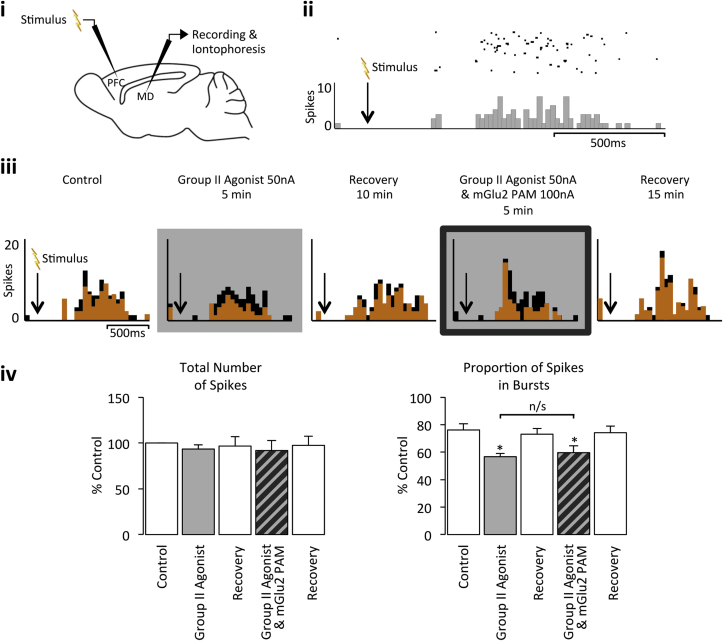
The mediodorsal thalamus (MD) likely plays an important role in cognition as it receives abundant afferent connections from the amygdala and prefrontal cortex (PFC). Indeed, disturbed activity within the MD is thought to precipitate cognitive deficits associated with schizophrenia. As compounds acting at the Group II metabotropic glutamate (mGlu) receptors (subtypes mGlu2/mGlu3) have efficacy in animal models of schizophrenia, we investigated whether a Group II agonist and an mGlu2 positive allosteric modulator (PAM) could modulate MD activity. Extracellular single-unit recordings were made in vivo from MD neurones in anaesthetised rats. Responses were elicited by electrical stimulation of the PFC and/or amygdala, with Group II compounds locally applied as required. The Group II agonist reduced inhibition evoked in the MD: an effect manifested as an increase in short-latency responses, and a decrease in long-latency burst-firing. This disinhibitory action of the Group II receptors in the MD represents a mechanism of potential therapeutic importance as increased inhibition in the MD has been associated with cognitive deficit-onset. Furthermore, as co-application of the mGlu2 PAM did not potentiate the Group II agonist effects in the MD, we suggest that the Group II disinhibitory effect is majority-mediated via mGlu3. This heterogeneity in Group II receptor thalamic physiology bears consequence, as compounds active exclusively at the mGlu2 subtype are unlikely to perturb maladapted MD firing patterns associated with cognitive deficits, with activity at mGlu3 receptors possibly more appropriate. Indeed, polymorphisms in the mGlu3, but not the mGlu2, gene have been detected in patients with schizophrenia.
Keywords: Burst firing; Egluetad (PubChem CID 156665); Mediodorsal thalamus; Metabotropic glutamate receptor; N-(4-(2-methoxyphenoxy)phenyl)-N-(2,2,2-trifluoroethylsulfonyl)pyrid-3-ylmethylamine (PubChem CID 9825084); N-Methylaspartate (PubChem CID 22880); Schizophrenia; Synaptic inhibition.
Copyright © 2015 The Authors. Published by Elsevier Ltd.. All rights reserved.
Figures


Similar articles
-
Actions of Xanthurenic acid, a putative endogenous Group II metabotropic glutamate receptor agonist, on sensory transmission in the thalamus.Neuropharmacology. 2013 Mar;66:133-42. doi: 10.1016/j.neuropharm.2012.03.009. Epub 2012 Apr 2. Neuropharmacology. 2013. PMID: 22491023
-
Astrocytes modulate thalamic sensory processing via mGlu2 receptor activation.Neuropharmacology. 2017 Jul 15;121:100-110. doi: 10.1016/j.neuropharm.2017.04.019. Epub 2017 Apr 14. Neuropharmacology. 2017. PMID: 28416443 Free PMC article.
-
Group II metabotropic glutamate receptor (mGlu2 and mGlu3 ) roles in thalamic processing.Br J Pharmacol. 2022 Apr;179(8):1607-1619. doi: 10.1111/bph.15640. Epub 2021 Sep 15. Br J Pharmacol. 2022. PMID: 34355803
-
The mediodorsal thalamus as a higher order thalamic relay nucleus important for learning and decision-making.Neurosci Biobehav Rev. 2015 Jul;54:76-88. doi: 10.1016/j.neubiorev.2015.03.001. Epub 2015 Mar 7. Neurosci Biobehav Rev. 2015. PMID: 25757689 Review.
-
Targeting metabotropic glutamate receptors for novel treatments of schizophrenia.Mol Brain. 2017 Apr 26;10(1):15. doi: 10.1186/s13041-017-0293-z. Mol Brain. 2017. PMID: 28446243 Free PMC article. Review.
Cited by
-
Cystine/Glutamate Antiporter and Aripiprazole Compensate NMDA Antagonist-Induced Dysfunction of Thalamocortical L-Glutamatergic Transmission.Int J Mol Sci. 2018 Nov 19;19(11):3645. doi: 10.3390/ijms19113645. Int J Mol Sci. 2018. PMID: 30463253 Free PMC article.
-
Thalamo-cortical neural mechanism of sodium salicylate-induced hyperacusis and anxiety-like behaviors.Commun Biol. 2024 Oct 18;7(1):1346. doi: 10.1038/s42003-024-07040-5. Commun Biol. 2024. PMID: 39420035 Free PMC article.
-
The hidden link: Investigating functional connectivity of rarely explored sub-regions of thalamus and superior temporal gyrus in Schizophrenia.Transl Neurosci. 2024 Dec 11;15(1):20220356. doi: 10.1515/tnsci-2022-0356. eCollection 2024 Jan 1. Transl Neurosci. 2024. PMID: 39669226 Free PMC article.
-
Lysergic acid diethylamide differentially modulates the reticular thalamus, mediodorsal thalamus, and infralimbic prefrontal cortex: An in vivo electrophysiology study in male mice.J Psychopharmacol. 2021 Apr;35(4):469-482. doi: 10.1177/0269881121991569. Epub 2021 Mar 1. J Psychopharmacol. 2021. PMID: 33645311 Free PMC article.
-
The role of thalamic group II mGlu receptors in health and disease.Neuronal Signal. 2022 Nov 15;6(4):NS20210058. doi: 10.1042/NS20210058. eCollection 2022 Dec. Neuronal Signal. 2022. PMID: 36561092 Free PMC article. Review.
References
-
- Adams D.H., Kinon B.J., Baygani S., Millen B.A., Velona I., Kollack-Walker S., Walling D.P. A long-term, phase 2, multicenter, randomized, open-label, comparative safety study of pomaglumetad methionil (LY2140023 monohydrate) versus atypical antipsychotic standard of care in patients with schizophrenia. BMC Psychiatry. 2013;13:143. - PMC - PubMed
-
- Aghajanian G.K., Marek G.J. Serotonin, via 5-HT2A receptors, increases EPSCs in layer V pyramidal cells of prefrontal cortex by an asynchronous mode of glutamate release. Brain Res. 1999;825(1–2):161–171. - PubMed
-
- Andrews J., Wang L., Csernansky J.G., Gado M.H., Barch D.M. Abnormalities of thalamic activation and cognition in schizophrenia. Am. J. Psychiatry. 2006;163(3):463–469. - PubMed
-
- Barbaresi P., Spreafico R., Frassoni C., Rustioni A. GABAergic neurons are present in the dorsal column nuclei but not in the ventroposterior complex of rats. Brain Res. 1986;382(2):305–326. - PubMed
Publication types
MeSH terms
Substances
Grants and funding
LinkOut - more resources
Full Text Sources
Other Literature Sources
Research Materials
Miscellaneous
