. 2015 Feb 20;6(5):2779-93.
doi: 10.18632/oncotarget.2995.

Sema6A and Mical1 control cell growth and survival of BRAFV600E human melanoma cells

Affiliations

Sema6A and Mical1 control cell growth and survival of BRAFV600E human melanoma cells

Rossella Loria et al. Oncotarget. .

Abstract

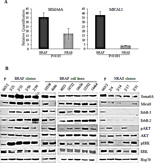
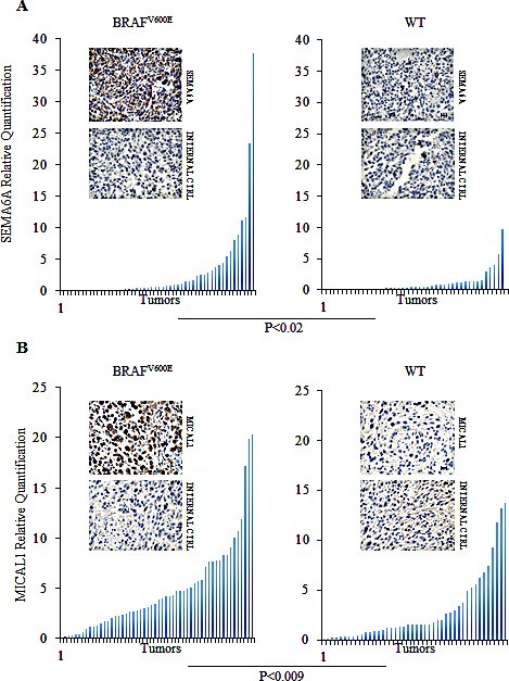
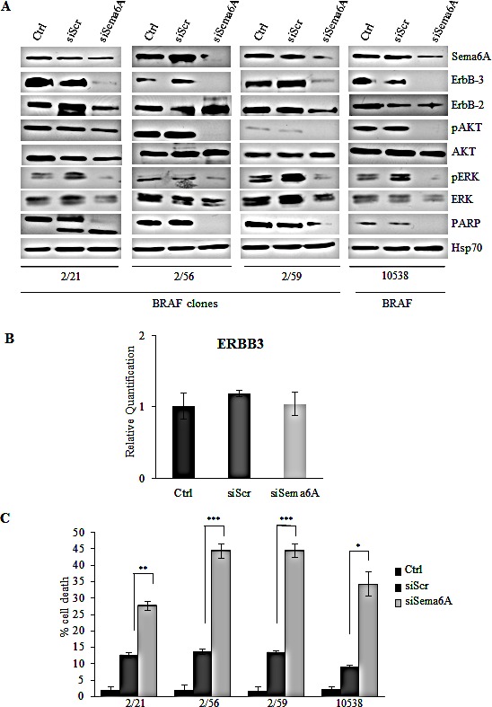
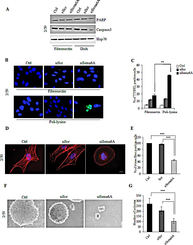
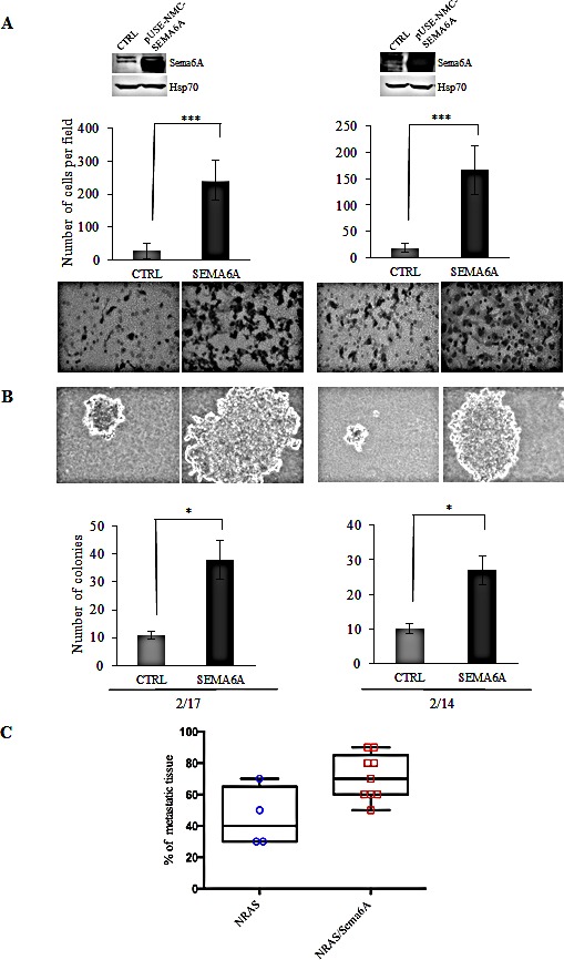
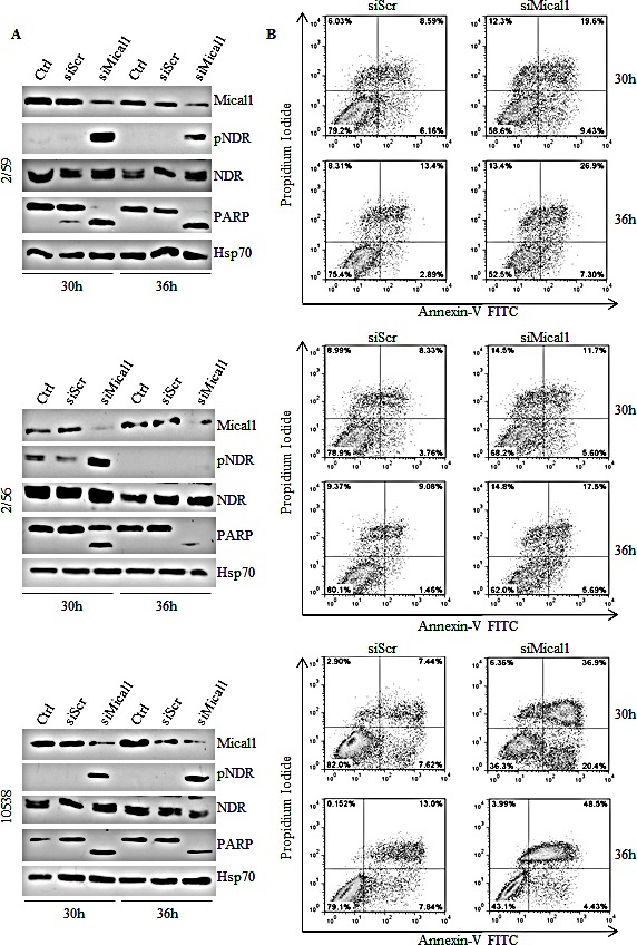
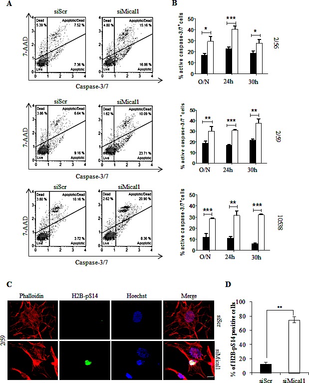
We used whole genome microarray analysis to identify potential candidate genes with differential expression in BRAFV600E vs NRASQ61R melanoma cells. We selected, for comparison, a peculiar model based on melanoma clones, isolated from a single tumor characterized by mutually exclusive expression of BRAFV600E and NRASQ61R in different cells. This effort led us to identify two genes, SEMA6A and MICAL1, highly expressed in BRAF-mutant vs NRAS-mutant clones. Real-time PCR, Western blot and immunohistochemistry confirmed preferential expression of Sema6A and Mical1 in BRAFV600E melanoma. Sema6A is a member of the semaphorin family, and it complexes with the plexins to regulate actin cytoskeleton, motility and cell proliferation. Silencing of Sema6A in BRAF-mutant cells caused cytoskeletal remodeling, and loss of stress fibers, that in turn induced cell death. Furthermore, Sema6A depletion caused loss of anchorage-independent growth, inhibition of chemotaxis and invasion. Forced Sema6A overexpression, in NRASQ61R clones, induced anchorage-independent growth, and a significant increase of invasiveness. Mical1, that links Sema/PlexinA signaling, is also a negative regulator of apoptosis. Indeed, Mical-1 depletion in BRAF mutant cells restored MST-1-dependent NDR phosphorylation and promoted a rapid and massive NDR-dependent apoptosis. Overall, our data suggest that Sema6A and Mical1 may represent new potential therapeutic targets in BRAFV600E melanoma.


Conflict of interest statement

Disclosure of Potential Conflict of Interest

No potential conflicts of interest were disclosed

Conflict of interest

No potential conflicts of interest were disclosed

Figures

Figure 1
Figure 1. Sema6A and Mical1 are highly expressed in BRAFV600E compared to NRASQ61R melanoma
(A) Average expression of SEMA6A and MICAL1 mRNA examined by qRT-PCR±SD. (B) Total cell lysates from cell line 665/2, from BRAF and NRAS clones isolated from 665/2 and from seven BRAF-mutant cell lines were analyzed by WB for expression of Sema6A, Mical1, ErbB3, ErbB2, total and pAkt, total and pERK, and Hsp70.
Figure 2
Figure 2. Sema6A and Mical1 are highly expressed in vivo in B-RAFV600E compared to WT melanomas
(A-B) Expression of SEMA6A and MICAL1 mRNA was examined by qRT-PCR on lymph node biopsies from patients carrying BRAFV600E or WT tumors (left and right graphs). Representative Sema6A and Mical1 IHC on sections derived from same samples. Secondary antibody was used as internal control. Scale bar 30 μm.
Figure 3
Figure 3. Interference for Sema6A induces cells death, and inhibits PI3K pathway
(A) siSema6A/BRAFV600E clones and primary melanoma cell line 10538 were analyzed by WB for expression of Sema6A, ErbB-3, ErbB-2, P-AKT, P-ERK1/2, PARP, and Hsp70. (B) Average expression of ErbB-3 mRNA examined by qRT-PCR±SD in Control, siScr, and siSema6A in clone 2/59. (C) Cell death of siSema6A/BRAFV600E cells was evaluated at 48 h post-transfection by Trypan blue exclusion from three independent experiments; bars ± S.D.
Figure 4
Figure 4. Interference with Sema6A expression in BRAF 2/59 cells induces cytoskeletal remodeling and inhibits anchorage-independent growth
(A) Control, siScr- or siSema6A/BRAFV600E cells were plated onto FN coated-dish or conventional culture-dish. Total cell lysates were analyzed by WB with PARP, Caspase3, and Hsp70 antibodies. (B) Tunel assay of BRAF/siSema6A cells plated on FN and/or poli-lysine. (C) Histogram reported percentage of tunel positive cells (P<0.004). (D) The cells, 48 h post-transfection, were plated on poly-l lysine coated slides, and stained with phalloidin-TRITC to show actin filaments. Scale bar is 10 μm. (E) Histogram reported percentage of stress fiber positive-cells. (F) Ctrl, siScr and siSema6A/BRAFV600E cells soft agar assay. (G) Histogram reported the number of colonies obtained in soft agar assay in Ctrl, siScr and siSema6A cells.
Figure 5
Figure 5. Sema6A controls invasion
(A) Overexpression of Sema6A by WB (upper part of the figure), and invasion assay (lower part of the figure), and (B) soft agar assay, of control and Sema6A/NRASQ61R overexpressing clones (2/14 and 2/17). (C) Box plot of in vivo skeletal tissues metastases analysis was obtained by software Prism version 6.0.
Figure 6
Figure 6. Silencing Mical1 induces apoptosis
(A) siMical-1/BRAFV600E cell lysates from 2/59 and 2/56 clones, and 10538 primary tumor were analyzed at the indicated time post-transfection for expression of Mical1, total and pNDR, and PARP. (B) Apoptosis was evaluated by Annexin-V/PI flow cytometric assay.
Figure 7
Figure 7. Silencing Mical1 induces Caspase 3/7 activation and H2B phosphorylation on Serine 14
(A) siMical-1/BRAFV600Ecells were analyzed with Muse™ Caspase-3/7 assay. (B) Caspase-3/7 activity after transfection with siScr (black bars) or siMICAL1 (white bars) is reported. (C) Cells plated on poly-l lysine coated slides, stained with phalloidin-TRITC, and anti-H2B-pS14 that co-stains with Hoechst of apoptotic nuclei of siMical-1/BRAFV600E cells. (D) Scale bar is 10 μm. Histogram reported the percentage of H2B-pS14 positive cells.

References

    1. Gray-Schopfer V, Wellbrock C, Marais R. Melanoma biology and new targeted therapy. Nature. 2007;445:851–7. - PubMed
    1. Chapman PB, Hauschild A, Robert C, Haanen JB Ascierto P, Larkin J, Dummer R, Garbe C, Testori A, Maio M, Hogg D, Lorigan P, Lebbe C, et al. Improved survival with vemurafenib in melanoma with BRAF V600E mutations. N Engl J Med. 2011;364:2507–16. - PMC - PubMed
    1. Menzies AM, Long GV. Dabrafenib and Trametinib, Alone and in Combination for BRAF-Mutant Metastatic Melanoma. Clin Cancer Res. 2014;20:2035–43. - PubMed
    1. Shi H, Hugo W, Kong X, Hong A, Koya RC, Moriceau G, Chodon T, Guo R, Johnson DB, Dahlman KB, Kelley MC, Kefford RF, Chmielowski B, et al. Acquired resistance and clonal evolution in melanoma during BRAF inhibitory therapy. Cancer Discov. 2014;4:80–93. - PMC - PubMed
    1. Nazarian R, Shi H, Wang Q, Kong X, Koya RC, Lee H, Chen Z, Lee MK, Attar N, Sazegar H, Chodon T, Nelson SF, McArthur G, et al. Melanomas acquire resistance to B-RAF(V600E) inhibition by RTK or N-RAS up-regulation. Nature. 2010;468:973–7. - PMC - PubMed

Publication types

MeSH terms