Review
. 2015 Feb 15:402:113-9.
doi: 10.1016/j.mce.2014.11.029. Epub 2015 Jan 8.

The sexual dimorphism of obesity

Affiliations
Review

The sexual dimorphism of obesity

Biff F Palmer et al. Mol Cell Endocrinol. .

Abstract

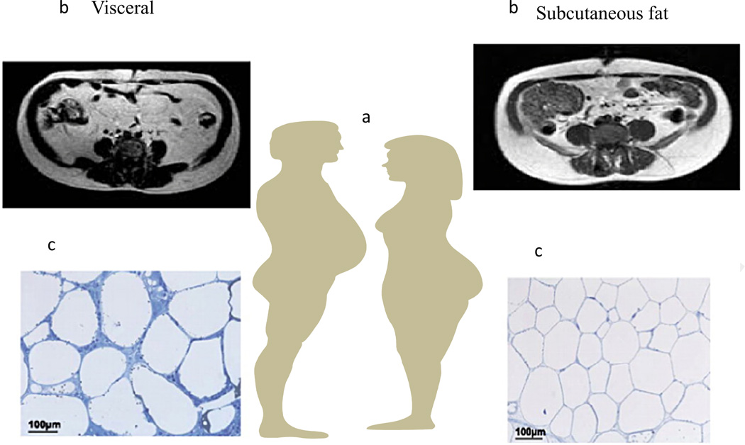
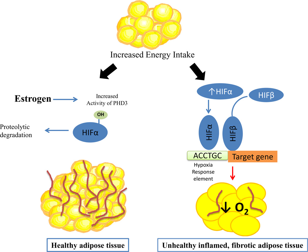
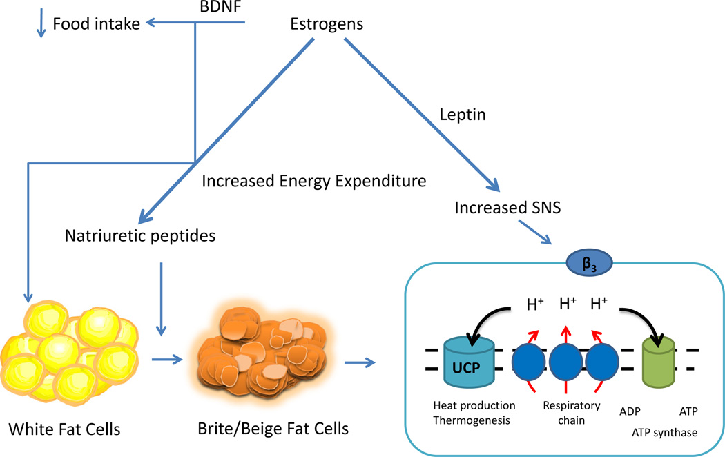
The NIH has recently highlighted the importance of sexual dimorphisms and has mandated inclusion of both sexes in clinical trials and basic research. In this review we highlight new and novel ways sex hormones influence body adiposity and the metabolic syndrome. Understanding how and why metabolic processes differ by sex will enable clinicians to target and personalize therapies based on gender. Adipose tissue function and deposition differ by sex. Females differ with respect to distribution of adipose tissues, males tend to accrue more visceral fat, leading to the classic android body shape which has been highly correlated to increased cardiovascular risk; whereas females accrue more fat in the subcutaneous depot prior to menopause, a feature which affords protection from the negative consequences associated with obesity and the metabolic syndrome. After menopause, fat deposition and accrual shift to favor the visceral depot. This shift is accompanied by a parallel increase in metabolic risk reminiscent to that seen in men. A full understanding of the physiology behind why, and by what mechanisms, adipose tissues accumulate in specific depots and how these depots differ metabolically by sex is important in efforts of prevention of obesity and chronic disease. Estrogens, directly or through activation of their receptors on adipocytes and in adipose tissues, facilitate adipose tissue deposition and function. Evidence suggests that estrogens augment the sympathetic tone differentially to the adipose tissue depots favoring lipid accumulation in the subcutaneous depot in women and visceral fat deposition in men. At the level of adipocyte function, estrogens and their receptors influence the expandability of fat cells enhancing the expandability in the subcutaneous depot and inhibiting it in the visceral depot. Sex hormones clearly influence adipose tissue function and deposition, determining how to capture and utilize their function in a time of caloric surfeit, requires more information. The key will be harnessing the beneficial effects of sex hormones in such a way as to provide 'healthy' adiposity.

Keywords: Estrogen, estrogen receptor alpha; Obesity; Sexual dimorphism; Subcutaneous fat depot; Visceral fat depot.


Figures

Fig. 1
Fig. 1
Approximately 80% of all body fat is in the subcutaneous depot and lies just under the skin primarily around the waist, in the subscapular area, and in the gluteal and femoral (thigh) areas. Visceral fat, accounting for 10–20% of total fat, is in the abdomen primarily in the omentum and mesentery but also in perirenal, gonadal, epicardial, and retroperitoneal depots. Visceral fat accounts for a higher percentage of total fat in men than in women. In men adipose tissue preferentially accumulates in the visceral depot while fat accumulation is primarily in the subcutaneous depot in women. The magnitude of this difference is amplified from late puberty to early adulthood as men develop the typical android body shape while women a more gynoid shape. Menopause is followed by redistribution of adipose tissue to the visceral depots leading to a more central or android shape in post-menopausal women who are not hormone replaced. The timing of these changes implicates involvement of sex hormones. Up to the transition through menopause, women tend to accrue adipose tissue preferentially in the subcutaneous depot due to its greater storage capacity, and the expandability of subcutaneous fat can be traced to a greater degree of hyperplasia of fat cells. Men accrue adipose tissue preferentially in the visceral depot, and the accumulation of excess fat in the visceral depot is primarily achieved by hypertrophy of fat cells. Once storage capacity is exceeded, visceral adipose tissue is characterized by fibrotic and inflamed adipose tissue which is highly correlated with the metabolic syndrome. (a) A cartoon depicting android and gynoid deposition of adipose tissue in males and females. (b) Representative coronal midsection MRI images of a BMI-matched male and female demonstrating fat distribution with the white matter depicting adipose tissues. (c) Representative histologic adipose tissue sections from subcutaneous or visceral adipose tissues. The subcutaneous adipose tissue has smaller more ‘plastic’ adipocytes whereas the visceral adipose tissue is characterized by larger adipocytes encased in fibrotic tissues.
Fig. 2
Fig. 2
When faced with chronic, excessive energy intake resulting in obesity, visceral adipose tissue (AT) becomes hypoxic due to increased adipocyte size and tissue mass which outpaces the supporting vascular supply resulting in inflamed and fibrotic adipose tissues which are co-localized with macrophages suggesting an immediate link between hypoxia and the inflammatory response. Both ERα and HIF-1 are related to fibrosis and inflammation in adipose tissue, albeit in opposite ways. ERα signaling improves adipose tissue function by decreasing adipose tissue inflammation and improving adipose tissue insulin sensitivity while HIF-1 worsens it through up-regulation of inflammatory mediators to include IL-6, NF-kB and TNFα and markers of fibrosis such as Col-6. It has recently been demonstrated E2/ERα regulates HIF-1 activity in adipose tissues by promoting transcription of a specific prolyl hydroxylase domain enzyme (PHD3). Increased activity of PHD3 through hydroxylation targets HIF for ubiquitination and degradation, thus providing a mechanistic explanation for the protective effect of E2/ERα against the metabolic impact of HIF-1 activation in adipose tissue and contributing to the reduction in adipose tissue inflammation and fibrosis seen in female adipose tissues.
Fig. 3
Fig. 3
Estrogens protect against increased body adiposity/obesity through their effects to suppress appetite and increase energy expenditure. In addition to enhancing potency of other anorectic signals and decreasing the potency of orexigenic signals, ERα signaling enhances leptin-induced satiety and increased energy expenditure. Leptin also induces activation of peripheral sympathetic nerve activity leading to increased energy expenditure. Estrogens/ERα enhance the release of natriuretic peptides, ANP and BNP, which have been demonstrated to act on adipose tissues to facilitate ‘browning’ through upregulation of UCP-1 which uncouples oxidative phosphorylation causing an increase in thermogenesis and increased energy expenditure. Estrogens also upregulate BDNF which has been demonstrated to play a role in suppressing appetite and increasing energy expenditure through effects associated with browning of adipose tissue. Therefore, estrogens through their activation of ERα have a pleiotropic effect on energy expenditure by increasing leptin induced activation of SNS, by upregulating BDNF, and by increasing ANP/BNP which facilitate the transition from white adipocytes to brown which are metabolically more energetic.

References

    1. Abildgarrd JPA, Green CJ, Harder-Lauridsen NM, Solomon TP, Thomsen C, Juul A, et al. Menopause is associated with decreased whole body fat oxidation during exercise. Am. J. Physiol. Endocrinol. Metab. 2013;304:E1227–E1236. - PubMed
    1. Adler ES, Hollis JH, Clarke IJ, Grattan DR, Oldfield BJ. Neurochemical characterization and sexual dimorphism of projections from the brain to abdominal and subcutaneous white adipose tissue in the rat. J. Neurosci. 2012;32:15913–15921. - PMC - PubMed
    1. Arner P, Lithell H, Wahrenberg H, Bronnegard M. Expression of lipoprotein lipase in different human subcutaneous adipose tissue regions. J. Lipid Res. 1991;32:423–429. - PubMed
    1. Asarian L, Geary N. Sex differences in the physiology of eating. Am. J. Physiol. Regul. Integr. Comp. Physiol. 2013;305:R1215–R1267. - PMC - PubMed
    1. Barr SI, Janelle KC, Prior JC. Energy intakes are higher during the luteal phase of ovulatory menstrual cycles. Am. J. Clin. Nutr. 1995;61:39–43. - PubMed