. 2015 Apr;11(2):242-53.
doi: 10.1007/s12015-014-9581-5.

Improved approach for chondrogenic differentiation of human induced pluripotent stem cells

Affiliations

Improved approach for chondrogenic differentiation of human induced pluripotent stem cells

Hossein Nejadnik et al. Stem Cell Rev Rep. 2015 Apr.

Abstract

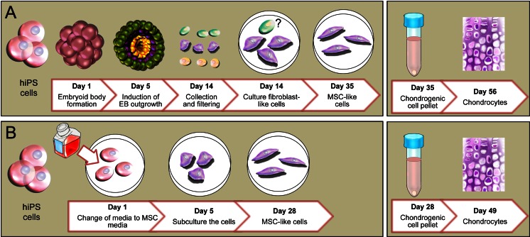
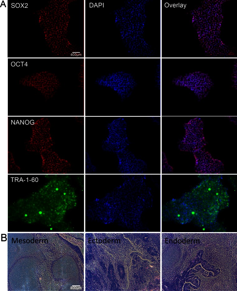
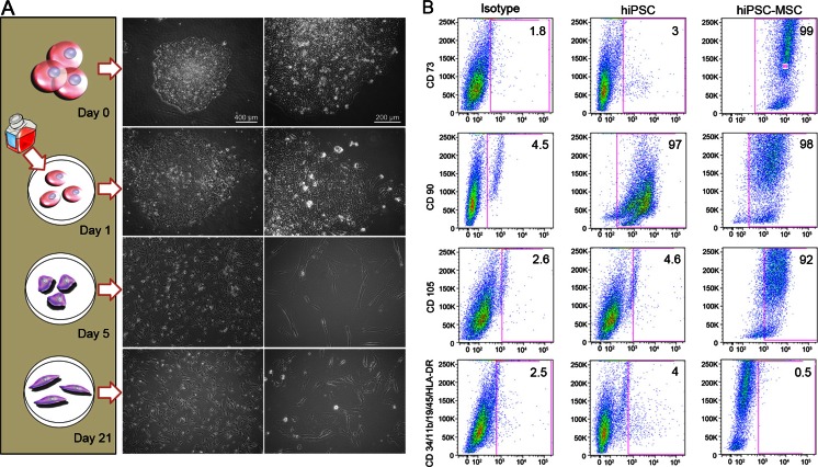
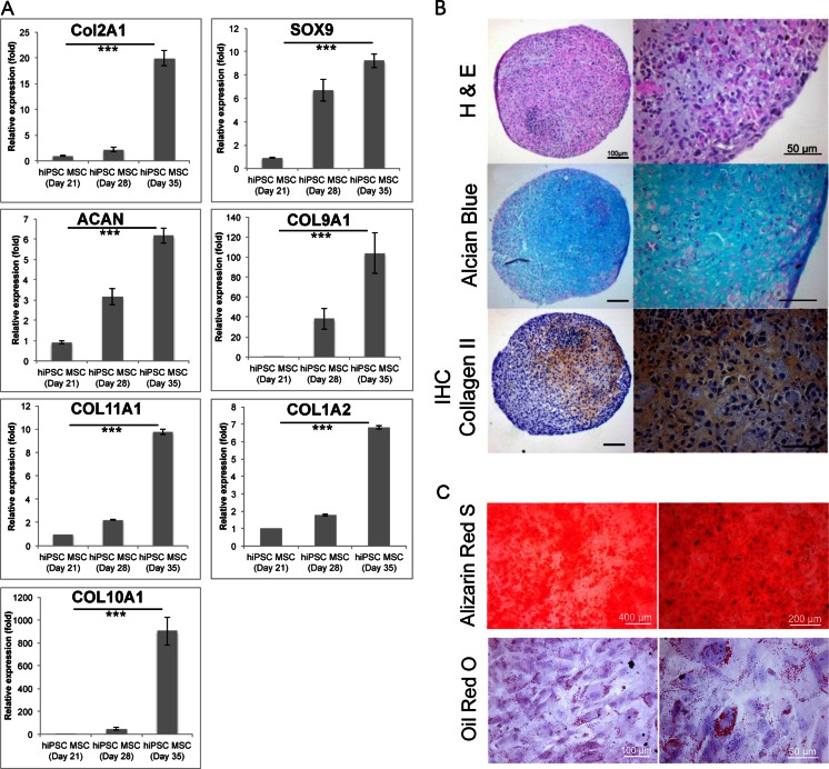
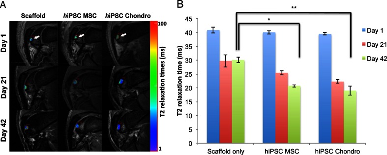
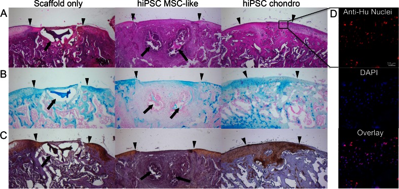
Human induced pluripotent stem cells (hiPSCs) have demonstrated great potential for hyaline cartilage regeneration. However, current approaches for chondrogenic differentiation of hiPSCs are complicated and inefficient primarily due to intermediate embryoid body formation, which is required to generate endodermal, ectodermal, and mesodermal cell lineages. We report a new, straightforward and highly efficient approach for chondrogenic differentiation of hiPSCs, which avoids embryoid body formation. We differentiated hiPSCs directly into mesenchymal stem /stromal cells (MSC) and chondrocytes. hiPSC-MSC-derived chondrocytes showed significantly increased Col2A1, GAG, and SOX9 gene expression compared to hiPSC-MSCs. Following transplantation of hiPSC-MSC and hiPSC-MSC-derived chondrocytes into osteochondral defects of arthritic joints of athymic rats, magnetic resonance imaging studies showed gradual engraftment, and histological correlations demonstrated hyaline cartilage matrix production. Results present an efficient and clinically translatable approach for cartilage tissue regeneration via patient-derived hiPSCs, which could improve cartilage regeneration outcomes in arthritic joints.


Figures

Fig. 1
Fig. 1
Chondrogenic differentiation of hiPSC. (a) Classical chondrogenic differentiation of hiPSCs via formation of embryoid bodies, outgrowth of endodermal (green), ectodermal (yellow) and mesodermal (red) cell lineages, selection of mesodermal cells, and induction of MSC and induction of chondrocytes. In this method hiPS cells were detached from matrigel coated dish and moved to ultra low attachment culture dish for 5 days to induce the EB formation, then EBs moved to plastic culture dish to select the hMSCs by collecting the outgrowing cells from EB (from day 5 to day 14) after collecting the attached fibroblast-like cells. These cells were cultured for 3 weeks in media containing FBS to prepare the hiPSC-MSCs (day 35 of differentiation). Then, hiPSC-MSCs were differentiated in a pellet culture system using serum free chondrogenic media for 3 weeks. (b) Embryoid body free method of direct differentiation of hiPSCs into hiPSC-MSCs, followed by chondrogenic differentiation. In embryoid body free method hiPSCs were cultured in matrigel coated dish and media was changed to hMSC media (DMEM supplemented with FBS) for 5 days to induce the hMSC differentiation (Day 5). Then, cells were detached and moved to a plastic culture dish for 4 passages to prepare the hiPSC-MSCs (Day 28). To differentiate the hiPSC-MSCs to chondrocytes, cells were used in pellet culture system using serum free chondrogenic media for 3 weeks
Fig. 2
Fig. 2
Pluripotency evaluation and teratoma formation of hiPSCs. (a) Immunofluorescence staining with DAPI counterstain demonstrating positive pluripotency markers NANOG, OCT4, SOX2, and TRA-1-60. (b) H&E stains of a representative hiPSC-derived teratoma confirm pluripotency of the hiPSCs with presence of all three germ layers, including ectoderm, mesoderm, and endoderm. (scale bar = 400 μm)
Fig. 3
Fig. 3
Morphology and Phenotypes of hiPSC-derived hiPSC-MSC cells. (a) Changes in cell morphology during hiPSC differentiation: Day 0: Dome-shaped hiPSC colony; Day 1: Changing the mTeSR1/E8 medium to hMSC medium leads to differentiation and out-growth of the cells from the colonies; Day 5: After sub-culture to uncoated and untreated culture flasks, the pre-differentiated cells attach to the polystyrene flask and start to form an elongated morphology. Day 21: At passage 4, cells show a spindle shape morphology, similar to hMSCs. (scale bar left panel = 400 and right panel 200 μm). (b) Flow cytometry analysis of surface markers of hiPSCs and hiPSC-MSCs at passage 4 shows positive hMSC surface markers of CD105, CD73, and CD90, and lack of CD45, CD34, CD14 or CD11b, CD19 and HLA-DR surface molecules according to the International Society for Cell Therapy (ISCT) criteria
Fig. 4
Fig. 4
Characterization of hiPSC-derived Chondrogenic Cells. (a) Relative gene expression of hiPSC-derived hiPSC-MSCs at day 21 (equal to day 0 of chondrogenic differentiation) and chondrogenic cell pellets at day 28 (equal to day 7 of chondrogenic differentiation) and 35 (equal to day 14 of chondrogenic differentiation), as determined by qPCR. Data are displayed as means and standard errors of triplicate experiments per sample. Cells at day 14 of chondrogenic differentiation show significantly increased gene expression of the hyaline chondrogenic markers COL2A1, COL9A1, COL11A1, SOX9, and aggrecan (ACAN) compared to hiPSC-MSCs. They also show an increased expression of COL1A2 and COL10A1 representative of fibro- and hypertrophic cartilage respectively. (*** indicates p < 0.001). (b) Histological evaluation of hiPSC-derived chondrogenic cell pellets at day 42 (equal to day 21 of chondrogenic differentiation); H&E stain shows chondrocytes and formation of a chondrogenic matrix. Alcian blue stain demonstrates positive glycosaminoglycan production, and immunohistochemistry shows positive stains for Collagen type II. (left 100 μm, right 50 μm). (c) Histological evaluation of hiPSC-MSCs osteogenic (upper panel) and adipogenic (lower panel) differentiation. Alizarin Red S staining used for osteogenic differentiation evaluation and Oil Red O staining used to assess the adipogenic differentiation of hiPSC-MSCs after 3 weeks of differentiation
Fig. 5
Fig. 5
In vivo engraftment of hiPSC-derived MSCs and Chondrogenic Cells. (a) Sagittal T2-weighted MR images of implants of scaffold only, hiPSC-derived MSCs, and hiPSC-derived chondrogenic cells in osteochondral defects of the distal femurs of rat knee joints. Superimposed T2 relaxation time maps show decreasing T2 values of transplanted cells, but not scaffold only, over time. (b) Corresponding quantitative measures of T2-relaxation times of cell transplants and scaffold only. Data are displayed as means and SE of triplicate experiments. (* and ** indicates p < 0.05 and p < 0.01 respectively)
Fig. 6
Fig. 6
Histological evaluation of hiPSC-derived MSCs and chondrogenic cells implanted in rat knee joints. (a) H&E stain of chondrogenic differentiated hiPSC-derived MSCs shows persistent defect after transplantation of scaffold only and engraftment of cell implants with defect remodeling. (b) Alcian blue stain demonstrates no glycosaminoglycan (GAG) production of scaffold only, mildly positive GAG production of hiPSC-derived MSCs and markedly positive GAG production of chondrogenic cells. (c) Collagen II immunohistochemistry shows no production of Collagen type II in scaffold only and MSC transplants, but markedly positive Collagen type II production after transplantation of chondrogenic cells. (Arrow heads display the borders of the defect and the complete arrows show the residual of scaffold; scale bar is equal to 500 μm). (d) Human anti-nuclear specific immunofluorescent stain shows presence and long term viability of the human cells in the repaired tissue (Scale bar is equal to 50 μm)

Similar articles

Cited by

References

    1. Egloff C, Hugle T, Valderrabano V. Biomechanics and pathomechanisms of osteoarthritis. Swiss Medical Weekly. 2012;142:w13583. - PubMed
    1. Elders MJ. The increasing impact of arthritis on public health. The Journal of Rheumatology. Supplement. 2000;60:6–8. - PubMed
    1. Goldring MB, Goldring SR. Osteoarthritis. Journal of Cellular Physiology. 2007;213:626–634. doi: 10.1002/jcp.21258. - DOI - PubMed
    1. Kock L, van Donkelaar CC, Ito K. Tissue engineering of functional articular cartilage: the current status. Cell and Tissue Research. 2012;347:613–627. doi: 10.1007/s00441-011-1243-1. - DOI - PMC - PubMed
    1. Mafi R, Hindocha S, Mafi P, Griffin M, Khan WS. Sources of adult mesenchymal stem cells applicable for musculoskeletal applications - a systematic review of the literature. Open Orthopaedica Journal. 2011;5(Suppl 2):242–248. doi: 10.2174/1874325001105010242. - DOI - PMC - PubMed

Publication types

MeSH terms