. 2015 Jan 13;10(2):204-15.
doi: 10.1016/j.celrep.2014.12.027. Epub 2015 Jan 8.

Nucleosome contact triggers conformational changes of Rpd3S driving high-affinity H3K36me nucleosome engagement

Affiliations

Nucleosome contact triggers conformational changes of Rpd3S driving high-affinity H3K36me nucleosome engagement

Chun Ruan et al. Cell Rep. .

Abstract

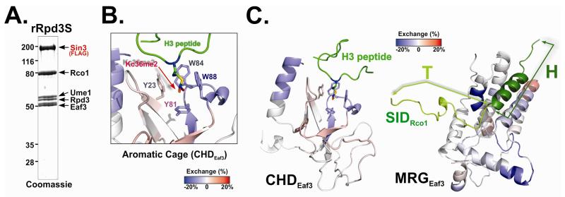
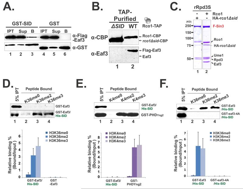
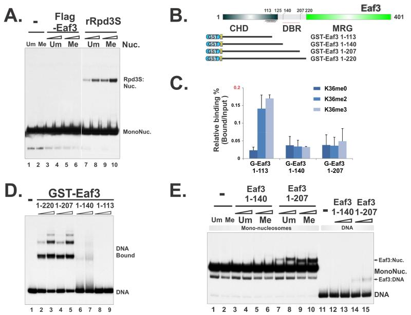
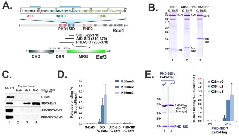
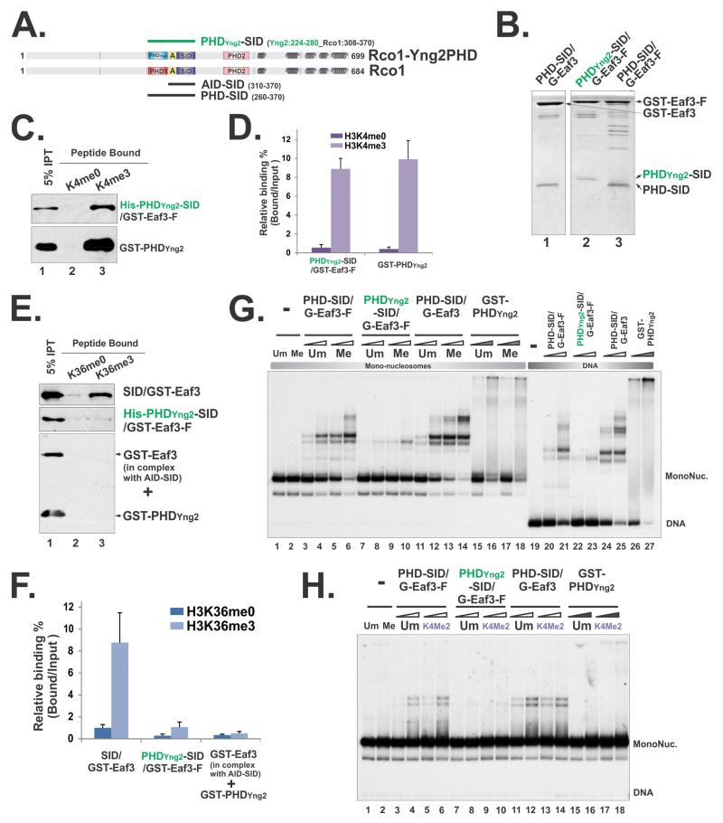
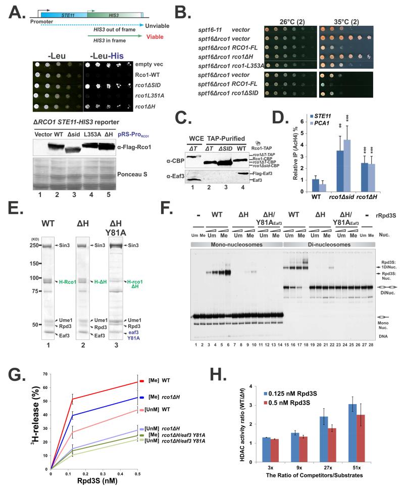
The Rpd3S histone deacetylase complex utilizes two subunits, Eaf3 and Rco1, to recognize nucleosomes methylated at H3K36 (H3K36me) with high affinity and strong specificity. However, the chromobarrel domain of Eaf3 (CHD) that is responsible for H3K36me recognition only binds weakly and with little specificity to histone peptides. Here, using deuterium exchange mass spectrometry (DXMS), we detected conformational changes of Rpd3S upon its contact with chromatin. Interestingly, we found that the Sin3-interacting domain of Rco1 (SID) allosterically stimulates preferential binding of Eaf3 to H3K36-methylated peptides. This activation is tightly regulated by an autoinhibitory mechanism to ensure optimal multivalent engagement of Rpd3S with nucleosomes. Lastly, we identified mutations at the interface between SID and Eaf3 that do not disrupt complex integrity but severely compromise Rpd3S functions in vitro and in vivo, suggesting that the nucleosome-induced conformational changes are essential for chromatin recognition.


Figures

Figure 1
Figure 1. Rpd3S undergoes conformation changes upon Rpd3S contact with nucleosomes
(A) Coomassie staining of the recombinant Rpd3S complex used in DXMS experiments. (B-C) Changes in the deuteration levels of Eaf3 upon Rpd3S binding to nucleosomes suggest conformational changes of Rpd3S; (B) Deuterium exchange results were mapped to 3D structure of the chromo domain of Eaf3 (PDB-2K3X). Blue color indicates slower deuterium exchange rates upon Rpd3S contact with nucleosomes, while red areas represent increased exchanges; Four aromatic residues that form the methyl-lysine binding pocket were labeled; (C) A zoom-out view of the deuterium exchange results of CHD (PDB: 2K3X) (Left) and MRG/SID (a molecular model based on PDB-2LKM using SWISS-MODEL)(the right panel). SID is represented in cartoon and green. The helix region of SID (dark green) is defined as the “H” region and the turn region (light green) is referred to as “T”. See also Figure S1.
Figure 2
Figure 2. Eaf3 can be allosterically activated to recognize H3K36me
(A-C) SID is required for incorporation of Eaf3 in Rpd3S; (A) GST-SID interacts with Flag-Eaf3 in vitro as shown by GST pull-down experiments. “IPT”-Input, “Sup”-Supernatant, “B”-Bound to beads; (B) Western blots of native Rpd3S that were TAP-purified from yeast strains YCR353(ΔSID) and YBL583(WT); (C) Coomassie staining of recombinant Rpd3S complexes purified from insect cells. (D-F) SID stimulates Eaf3 to recognize H3K36me preferentially. Histone peptide pull-down assays were performed using indicated proteins or protein complexes. The upper panels in each figures show representative western blot results, while the lower panels display quantification of the western results based on at least four independent experiments. Data are represented as mean ± SEM; (D) SID increases the binding of Eaf3 to H3K36 methylated peptides; (E) SID/Eaf3 heterodimer does not bind to H3K4 methylated peptides. The PHD domain of Yng2 was used as a positive control; (F) The elevated binding of SID/Eaf3 to H3K36 methylated peptides relies on the aromatic cage of Eaf3 CHD. pBL1290 was used to purify the cage mutant of SID/Eaf3, in which all four aromatic residues were mutated to alanine (GST-eaf3-4A). See also Figure S2.
Figure 3
Figure 3. DNA and histone binding abilities of Eaf3 are self-contained
(A) Full-length Eaf3 protein, purified from an insect-cell system, does not bind to nucleosomes and DNA. (B) Constructs for mapping Eaf3 DNA-binding regions and the histone H3K36me-binding subunit. (C) The binding of Eaf3 to histone H3K36me is auto-inhibited; Histone peptide binding assays were quantified based on three repeats. Data are represented as mean ± SEM. (D) The region of 140-207 of Eaf3 is a potential DNA-Binding Region. EMSA assay using 32P labeled 196-1X probe and GST-fused Eaf3 truncations. (E) The Eaf3 truncations that include CHD and DBR can weakly bind to nucleosomes as measured by EMSA using mono-nucleosome substrates. * indicates partially disintegrated nucleosomes, likely to be hexasomes. See also Figure S3 and S4.
Figure 4
Figure 4. SID-induced Eaf3 activation is controlled by an auto-inhibition mechanism
(A). An illustration of domain structures in Eaf3 and Rco1. (B) Coomassie staining of tandem purified Eaf3-Rco1 heterodimers. (C) Histone peptide pull-down assay show that AID suppresses the SID-mediated activation of Eaf3. (D) Quantification of (C) based on three repeats. Data are represented as mean ± SEM. (E) Coomassie staining of tandem purified AID mutated PHD-SID/Eaf3 heterodimers (left). Histone peptide binding assay (right). See also Figure S4 and S5.
Figure 5
Figure 5. PHD-SID/Eaf3 is the minimal nucleosome-binding module of Rpd3S
(A) The binding Rco1/Eaf3 heterodimers were tested in EMSA using mono-nucleosome substrates. (B) EMSA using DNA alone. (C-D) The DBR and the aromatic pocket of CHD are required for the binding of PHD-SID/Eaf3 heterodimers to nucleosomes; (C) Coomassie staining of tandem purified wild type and mutant PHD-SID/Eaf3 heterodimers; Plasmids pBL1291 and pBL1296 were used to purify PHD-SID/eaf3-Y81A and PHD-SID/ eaf3ΔDBR (116-206) respectively; (D) EMSA using mono-nucleosome substrates. See also Figure S4 and S6.
Figure 6
Figure 6. Perturbation of PHD domain within the minimal nucleosome binding module compromised its nucleosome engagement
(A) A schematic illustration of the PHDYng2-SID/Eaf3 construct. “A” framed in the yellow box represents the AID domain. Amino acids that were included in the hybrid protein were indicated by the residue numbers behind each constructs. (B) Coomassie staining of tandem purified hybrid Rco1-Eaf3 heterodimers. (C) Histone peptide pull-down using unmethylated and methylated H3K4 peptides. (D) Quantification of (C) based on three repeats. Data are represented as mean ± SEM. (E) Histone peptide pull-down using unmethylated and methylated H3K36 peptides. The low level binding of PHDYng2-SID/Eaf3 was likely due to PHDYng2-somehow slightly compromises AID function, because when AID-SID/Eaf3 heterodimers and GST-PHDYng2 were mixed together, no binding was detected. (F) Quantification of (E) based on three repeats. (G) EMSA using mono-nucleosome substrates that unmethylated or tri-methylated at H3K36 and DNA. Two concentrations of heterodimers were 15pM, 30pM respectively and indicated as open triangles. 3.2μM and 6.4μM of GST-PHDYng2 were used and labeled as filled triangles. (H) EMSA using mono-nucleosome substrates that unmethylated or di-methylated at H3K4. 1.6μM and 3.2μM of GST-PHDYng2 were used.
Figure 7
Figure 7. Conformational changes are essential for Rpd3S function
(A) Test cryptic transcription phenotype caused by Rco1 mutants in an STE11-HIS reporter strain (YCR239). The bottom panel: Western blot shows that all mutant proteins are expressed at similar levels. (B) Test Rco1 function in FACT mutants. Plasmids carrying wild type or mutant RCO1 under the control of its native promoter (parental vector pBL1114) were transformed into YBL823 (spt16-11 ΔRCO1). The resulting strains were subjected to spotting assays and grown at semi-permissive temperature. (C) Deletion of the “T” region (ΔT) results in the loss of Eaf3 from Rpd3S in vivo. TAP purified Rpd3S complexes were subjected to western blot to monitor the association of Eaf3 with Rpd3S. Noted that wild type Rco1 strain containing a Flag-Eaf3 (YBL768), therefore the Eaf3 bands as detected by a polyclonal antibody against Eaf3 migrates slower than untagged version (Lane 1). (D) ChIP assay using an antibody against AcH4 show that disruption of SID/Eaf3 interaction interface results in increased acetylation levels at coding regions of the STE11 and PCA1 gens. IP efficiency of each gene was normalized to AcH4 IP efficiency at the Y region (a gene desert on Chromosome 6 which serves as an internal control). Data are represented as mean ± SEM, N>3, *(P<0.05) **(P<0.01), ***(P<0.001) based on 2-tailed Student T-Test. (E-H) Deletion of the “H” region (ΔH) does not disrupt complex integrity but compromises Rpd3S functions in vitro; (E) Coomassie staining of Rpd3S ΔH mutants; (F) EMSA assays using mono- and di-nucleosomes; (G) Nucleosome-based HDAC assay for indicated Rpd3S complexes. The deacetylation activity, which is indicated by the amount of free 3H release, was plotted against the concentration of Rpd3S; (H) Defects caused by ΔH are more severe under stringent competition. Increasing amount of competitors (DNA and HeLa oligonucleosomes) were added into each HDAC reactions as shown in (G), The ratio of the HDAC activity of wild type Rpd3S over the ΔH mutant was shown as a function of the amount of competitors.

Similar articles

Cited by

References

    1. Biswas D, Takahata S, Stillman DJ. Different Genetic Functions for the Rpd3(L) and Rpd3(S) Complexes Suggest Competition Between NuA4 and Rpd3(S) Mol Cell Biol. 2008 - PMC - PubMed
    1. Burns-Hamuro LL, Hamuro Y, Kim JS, Sigala P, Fayos R, Stranz DD, Jennings PA, Taylor SS, Woods VL., Jr. Distinct interaction modes of an AKAP bound to two regulatory subunit isoforms of protein kinase A revealed by amide hydrogen/deuterium exchange. Protein Sci. 2005;14:2982–2992. - PMC - PubMed
    1. Carrozza MJ, Li B, Florens L, Suganuma T, Swanson SK, Lee KK, Shia WJ, Anderson S, Yates J, Washburn MP, Workman JL. Histone H3 methylation by Set2 directs deacetylation of coding regions by Rpd3S to suppress spurious intragenic transcription. Cell. 2005;123:581–592. - PubMed
    1. Cheung V, Chua G, Batada NN, Landry CR, Michnick SW, Hughes TR, Winston F. Chromatin- and transcription-related factors repress transcription from within coding regions throughout the Saccharomyces cerevisiae genome. PLoS Biol. 2008;6:e277. - PMC - PubMed
    1. Drouin S, Laramee L, Jacques PE, Forest A, Bergeron M, Robert F. DSIF and RNA polymerase II CTD phosphorylation coordinate the recruitment of Rpd3S to actively transcribed genes. PLoS Genet. 2010;6:e1001173. - PMC - PubMed

Publication types

MeSH terms

LinkOut - more resources