Visualizing endocytotic pathways at transmission electron microscopy via diaminobenzidine photo-oxidation by a fluorescent cell-membrane dye
- PMID: 25578976
- PMCID: PMC4289848
- DOI: 10.4081/ejh.2014.2449
Visualizing endocytotic pathways at transmission electron microscopy via diaminobenzidine photo-oxidation by a fluorescent cell-membrane dye
Abstract
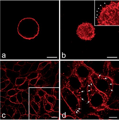
The endocytotic pathway involves a complex, dynamic and interacting system of intracellular compartments. PKH26 is a fluorescent dye specific for long-lasting cell membrane labelling which has been successfully used for investigating cell internalization processes, at either flow cytometry or fluorescence microscopy. In the present work, diaminobenzidine photo-oxidation was tested as a procedure to detect PKH26 dye at transmission electron microscopy. Our results demonstrated that DAB-photo-oxidation is a suitable technique to specifically visualise this fluorescent dye at the ultrastructural level: the distribution of the granular dark reaction product perfectly matches the pattern of the fluorescence staining, and the electron density of the fine precipitates makes the signal evident and precisely detectable on the different subcellular compartments involved in the plasma membrane internalization routes.
Figures


Similar articles
-
Photooxidation technology for correlated light and electron microscopy.J Microsc. 2009 Sep;235(3):322-35. doi: 10.1111/j.1365-2818.2009.03220.x. J Microsc. 2009. PMID: 19754726 Review.
-
Tracing nanoparticles and photosensitizing molecules at transmission electron microscopy by diaminobenzidine photo-oxidation.Micron. 2014 Apr;59:44-51. doi: 10.1016/j.micron.2013.12.007. Epub 2013 Dec 25. Micron. 2014. PMID: 24530364 Review.
-
FM dye photoconversion for visualizing synaptic vesicles by electron microscopy.Cold Spring Harb Protoc. 2012 Jan 1;2012(1):84-6. doi: 10.1101/pdb.prot067611. Cold Spring Harb Protoc. 2012. PMID: 22194271
-
Photoconversion of diaminobenzidine with different fluorescent neuronal markers into a light and electron microscopic dense reaction product.Microsc Res Tech. 1993 Jan 1;24(1):2-14. doi: 10.1002/jemt.1070240103. Microsc Res Tech. 1993. PMID: 7679591
-
Non-Specific Particle Formation During Extracellular Vesicle Labelling With the Lipophilic Membrane Dye PKH26.J Extracell Vesicles. 2025 May;14(5):e70079. doi: 10.1002/jev2.70079. J Extracell Vesicles. 2025. PMID: 40387660 Free PMC article.
Cited by
-
Nanoscopy for endosomal escape quantification.Nanoscale Adv. 2020 Oct 27;3(1):10-23. doi: 10.1039/d0na00454e. eCollection 2021 Jan 7. Nanoscale Adv. 2020. PMID: 36131870 Free PMC article. Review.
-
Ultrastructural histochemistry in biomedical research: Alive and kicking.Eur J Histochem. 2018 Nov 7;62(4):2990. doi: 10.4081/ejh.2018.2990. Eur J Histochem. 2018. PMID: 30418011 Free PMC article.
-
Isolation of Human Small Extracellular Vesicles and Tracking of their Uptake by Retinal Pigment Epithelial Cells In Vitro.Int J Mol Sci. 2020 May 27;21(11):3799. doi: 10.3390/ijms21113799. Int J Mol Sci. 2020. PMID: 32471212 Free PMC article.
-
Diaminobenzidine Photooxidation to Visualize Fluorescent Nanoparticles in Adhering Cultured Cells at Transmission Electron Microscopy.Methods Mol Biol. 2023;2566:333-343. doi: 10.1007/978-1-0716-2675-7_27. Methods Mol Biol. 2023. PMID: 36152264
-
Transmission electron microscopy for nanomedicine: novel applications for long-established techniques.Eur J Histochem. 2016 Dec 9;60(4):2751. doi: 10.4081/ejh.2016.2751. Eur J Histochem. 2016. PMID: 28076938 Free PMC article. Review.
References
-
- Panzeri D, Lavazza T, Malgaroli A. Advanced tracer techniques to monitor synaptic activity. Arch Ital Biol 2005; 143:157-68. - PubMed
-
- de Souza W, Sant’Anna C, Cunha-e-Silva NL. Electron microscopy and cytochemistry analysis of the endocytic pathway of pathogenic protozoa. Prog Histochem Cytochem 2009;44:67-124. - PubMed
-
- Bissig C, Johnson S, Gruenberg J. Studying lipids involved in the endosomal pathway. Methods Cell Biol 2012;108:19-46. - PubMed
MeSH terms
Substances
LinkOut - more resources
Full Text Sources
Other Literature Sources