

Optical coherence tomography in papilledema and pseudopapilledema with and without optic nerve head drusen
- PMID: 25579359
- PMCID: PMC4313495
- DOI: 10.4103/0301-4738.149136
Optical coherence tomography in papilledema and pseudopapilledema with and without optic nerve head drusen
Abstract
Aim: To compare the spectral domain optical coherence tomography (SD-OCT) findings of the optic disc and the peripapillary retina of patients with a true papilledema and pseudopapilledema with and without optic nerve head drusen (ONHD).
Study design: Retrospective Case Control Study.
Subjects and methods: Peripapillary retinal nerve fiber layer (PPRNFL) thickness as depicted by SD-OCT of 94 eyes of 66 patients with papilledema (30 eyes), pseudopapiledema (31 eyes), and normal controls (33 eyes) was analyzed. The mean RNFL thickness, total retinal thickness (TRT) at a superior and inferior edge of the disc and the quadrant wise topography of increased RNFL were compared in all three groups. Sensitivity, specificity, and area under the receiver operating characteristic curve (AROC) were calculated for all the parameters.
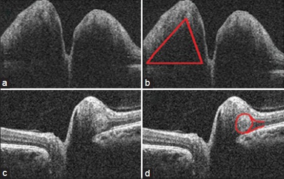
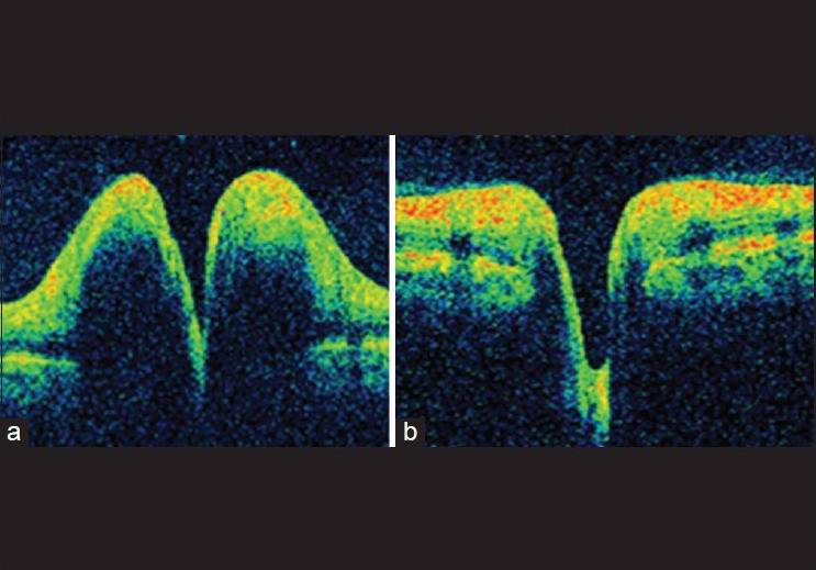
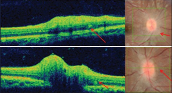
Results: The median RNFL thickness was 185.4 (129.5-349.3 μm), 122.3 (109-156.3 μm) and 91.62 ± 7 μm in papilledema, pseudopapilledema, and controls, respectively. Papilledema group had thicker PPRNFL in all quadrants except temporal quadrant. TRT was thicker in papilledema and pseudopapilledema compared to controls. ONHD could be directly visualized as high reflective clumps in the sub-retinal space or the RNFL in 30 eyes. Increased RNFL thickness in all four quadrants was noted 43.3% in papilledema and 9.7% in pseudopapilledema. Normal RNFL thickness in all four quadrants was noted in 0% in papilledema and 32.3% in pseudopapilledema. Nasal RNFL had the highest AROC (0.792) indicating high diagnostic ability to differentiate papilledema from pseudopapilledema.
Conclusion: SD-OCT can be used as a tool to differentiate between papilledema and pseudopapilledema.
Conflict of interest statement
Figures

References
-
- Kurz-Levin MM, Landau K. A comparison of imaging techniques for diagnosing drusen of the optic nerve head. Arch Ophthalmol. 1999;117:1045–9. - PubMed
-
- Mustonen E, Nieminen H. Optic disc drusen – A photographic study. I. Autofluorescence pictures and fluorescein angiography. Acta Ophthalmol (Copenh) 1982;60:849–58. - PubMed
-
- Vartin C V, Nguyen AM, Balmitgere T, Bernard M, Tilikete C, Vighetto A. Detection of mild papilloedema using spectral domain optical coherence tomography. Br J Ophthalmol. 2012;96:375–9. - PubMed
-
- Johnson LN, Diehl ML, Hamm CW, Sommerville DN, Petroski GF. Differentiating optic disc edema from optic nerve head drusen on optical coherence tomography. Arch Ophthalmol. 2009;127:45–9. - PubMed
-
- Lee KM, Woo SJ, Hwang JM. Differentiation of optic nerve head drusen and optic disc edema with spectral-domain optical coherence tomography. Ophthalmology. 2011;118:971–7. - PubMed
MeSH terms
Supplementary concepts
LinkOut - more resources
Full Text Sources
Other Literature Sources
Medical
