

Performance comparison of 1.5-T endorectal coil MRI with 3.0-T nonendorectal coil MRI in patients with prostate cancer
- PMID: 25579637
- PMCID: PMC4355101
- DOI: 10.1016/j.acra.2014.11.007
Performance comparison of 1.5-T endorectal coil MRI with 3.0-T nonendorectal coil MRI in patients with prostate cancer
Abstract
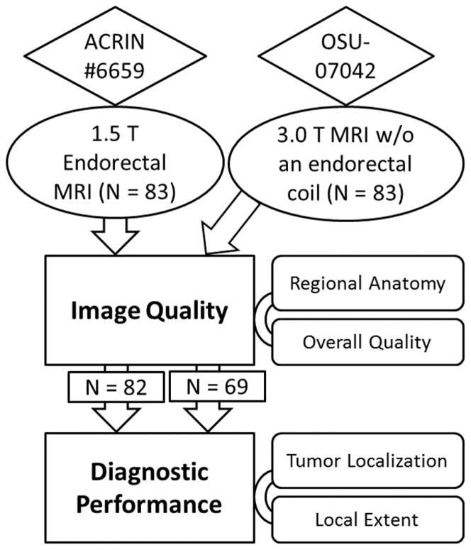
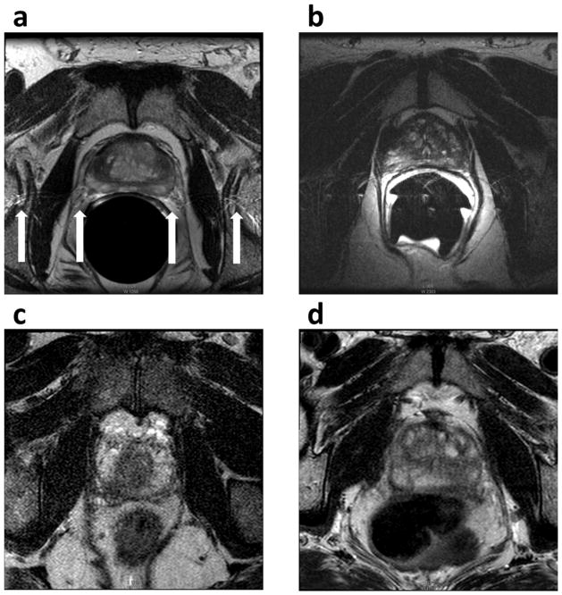
Rationale and objectives: To compare prostate morphology, image quality, and diagnostic performance of 1.5-T endorectal coil magnetic resonance (MR) imaging (MRI) and 3.0-T nonendorectal coil MRI in patients with prostate cancer.
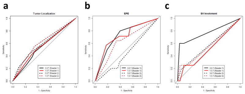
Materials and methods: MR images obtained of 83 patients with prostate cancer using 1.5-T MRI systems with an endorectal coil were compared to images collected from 83 patients with a 3.0-T MRI system. Prostate diameters were measured, and image quality was evaluated by one American Board of Radiology (ABR)-certified radiologist (reader 1) and one ABR-certified diagnostic medical physicist (reader 2). The likelihood of the presence of peripheral zone cancer in each sextant and local extent was rated and compared to histopathologic findings.
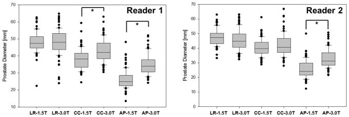
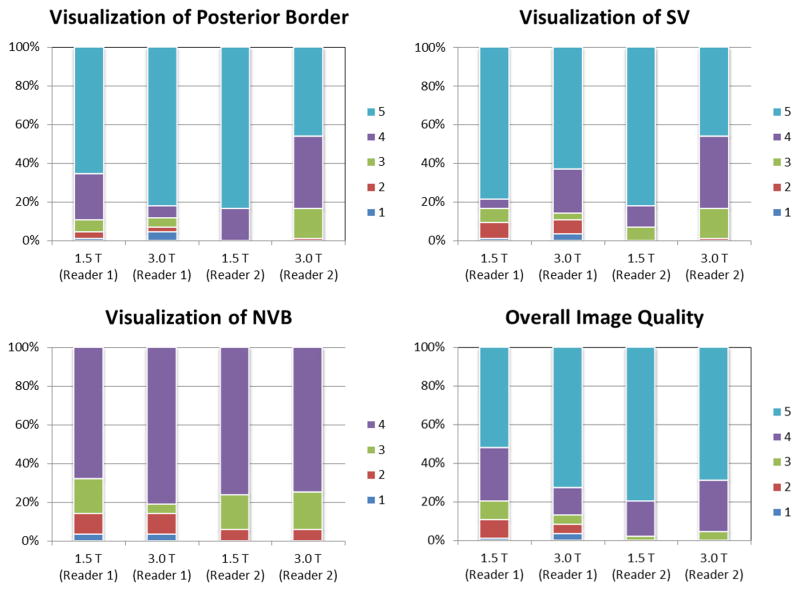
Results: Prostate anterior-posterior diameter measured by both readers was significantly shorter with 1.5-T endorectal MRI than with 3.0-T MRI. The overall image quality score difference was significant only for reader 1. Both readers found that the two MRI systems provided a similar diagnostic accuracy in cancer localization, extraprostatic extension, and seminal vesicle involvement.
Conclusions: Nonendorectal coil 3.0-T MRI provides prostate images that are natural in shape and that have comparable image quality to those obtained at 1.5 T with an endorectal coil, but not superior diagnostic performance. These findings suggest an opportunity exists for improving technical aspects of the 3.0-T prostate MRI.
Keywords: Prostate cancer; endorectal coil; image quality; magnetic resonance imaging; tumor localization; tumor staging.
Copyright © 2015 AUR. Published by Elsevier Inc. All rights reserved.
Figures
References
-
- Siegel R, Ma J, Zou Z, Jemal A. Cancer statistics, 2014. CA Cancer J Clin. 2014;64(1):9–29. - PubMed
-
- Mazaheri Y, Shukla-Dave A, Muellner A, Hricak H. MRI of the prostate: clinical relevance and emerging applications. J Magn Reson Imaging. 2011;33(2):258–74. - PubMed
-
- Hricak H, Choyke PL, Eberhardt SC, Leibel SA, Scardino PT. Imaging prostate cancer: a multidisciplinary perspective. Radiology. 2007;243(1):28–53. - PubMed
-
- Hricak H, White S, Vigneron D, et al. Carcinoma of the prostate gland: MR imaging with pelvic phased-array coils versus integrated endorectal--pelvic phased-array coils. Radiology. 1994;193(3):703–9. - PubMed
-
- Rajesh A, Coakley FV. MR imaging and MR spectroscopic imaging of prostate cancer. Magn Reson Imaging Clin N Am. 2004;12(3):557–79. - PubMed
Publication types
MeSH terms
Grants and funding
LinkOut - more resources
Full Text Sources
Other Literature Sources
Medical
