Review
. 2015 Apr:42:105-18.
doi: 10.1016/j.mam.2015.01.001. Epub 2015 Jan 8.

Proinsulin misfolding and endoplasmic reticulum stress during the development and progression of diabetes

Affiliations
Review

Proinsulin misfolding and endoplasmic reticulum stress during the development and progression of diabetes

Jinhong Sun et al. Mol Aspects Med. 2015 Apr.

Abstract

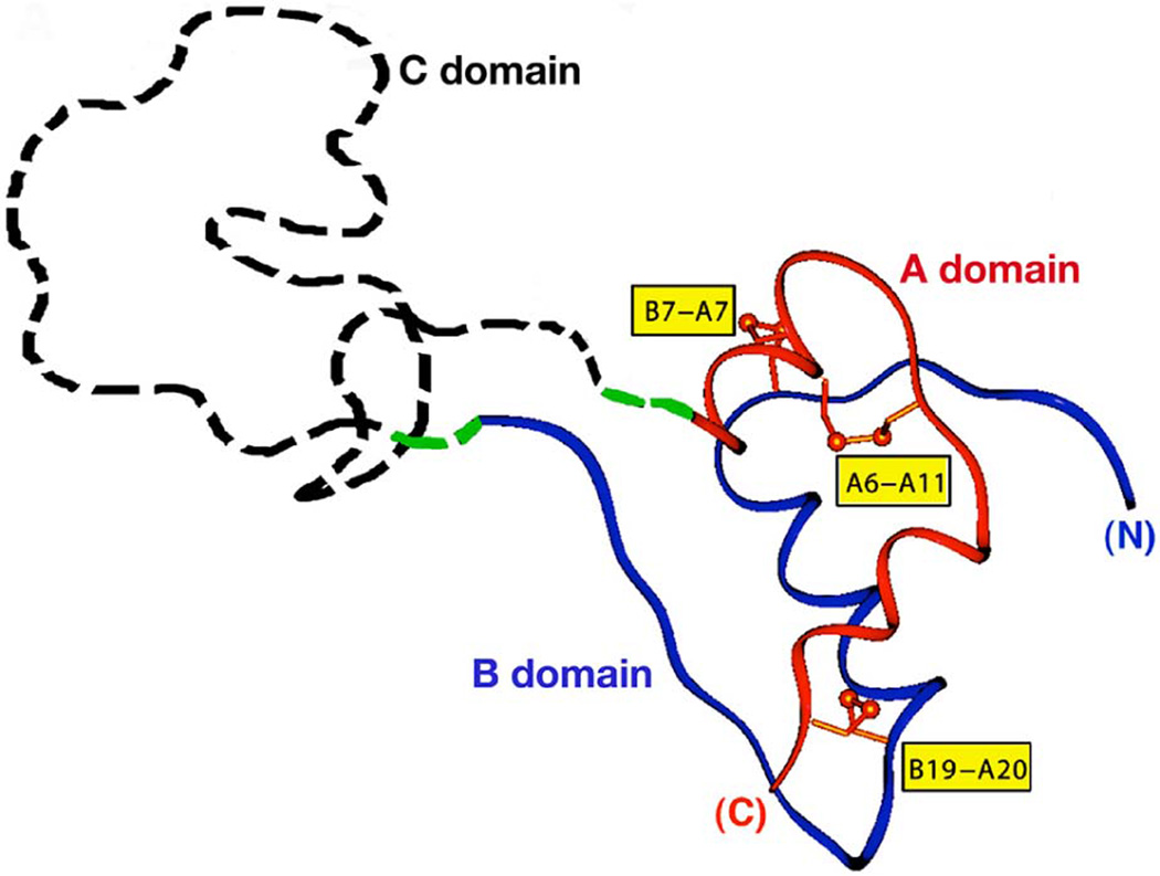
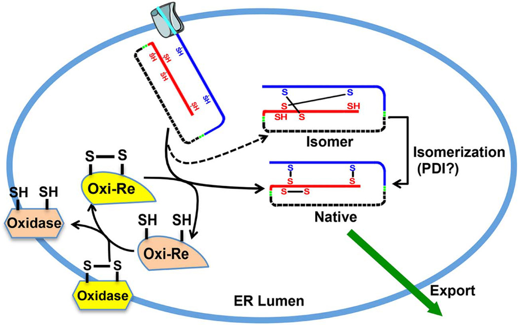
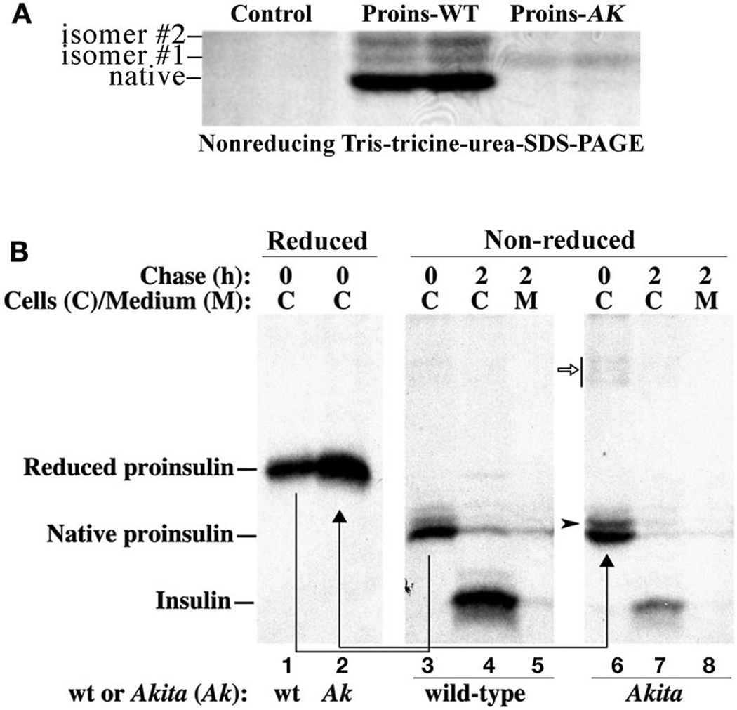
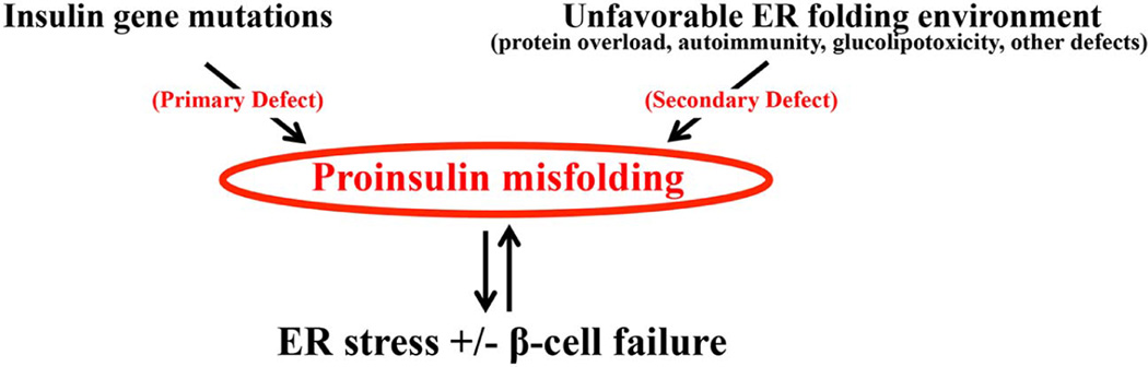
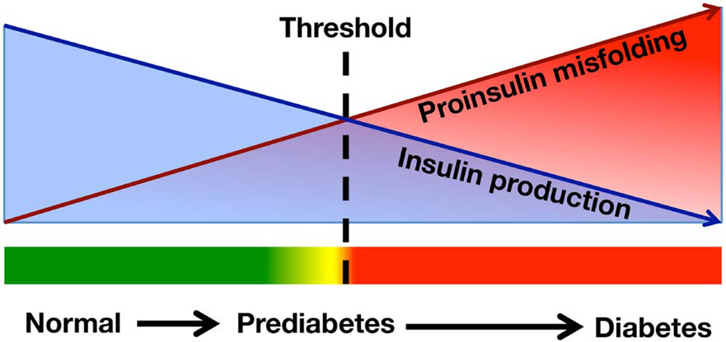
To maintain copious insulin granule stores in the face of ongoing metabolic demand, pancreatic beta cells must produce large quantities of proinsulin, the insulin precursor. Proinsulin biosynthesis can account for up to 30-50% of total cellular protein synthesis of beta cells. This puts pressure on the beta cell secretory pathway, especially the endoplasmic reticulum (ER), where proinsulin undergoes its initial folding, including the formation of three evolutionarily conserved disulfide bonds. In normal beta cells, up to 20% of newly synthesized proinsulin may fail to reach its native conformation, suggesting that proinsulin is a misfolding-prone protein. Misfolded proinsulin molecules can either be refolded to their native structure or degraded through ER associated degradation (ERAD) and autophagy. These degraded molecules decrease proinsulin yield but do not otherwise compromise beta cell function. However, under certain pathological conditions, proinsulin misfolding increases, exceeding the genetically determined threshold of beta cells to handle the misfolded protein load. This results in accumulation of misfolded proinsulin in the ER - a causal factor leading to beta cell failure and diabetes. In patients with Mutant INS-gene induced diabetes of Youth (MIDY), increased proinsulin misfolding due to insulin gene mutations is the primary defect operating as a "first hit" to beta cells. Additionally, increased proinsulin misfolding can be secondary to an unfavorable ER folding environment due to genetic and/or environmental factors. Under these conditions, increased wild-type proinsulin misfolding becomes a "second hit" to the ER and beta cells, aggravating beta cell failure and diabetes. In this article, we describe our current understanding of the normal proinsulin folding pathway in the ER, and then review existing links between proinsulin misfolding, ER dysfunction, and beta cell failure in the development and progression of type 2, type 1, and some monogenic forms of diabetes.

Keywords: Beta cell; Diabetes; Endoplasmic reticulum stress; Insulin biosynthesis; Proinsulin folding and misfolding.


Figures

Figure 1
Figure 1. Structure of proinsulin
Proinsulin is comprised sequentially of insulin B domain (blue), connecting C domain (which is disordered and therefore has no fixed structure), and insulin A domain (red). Six cysteines form three disulfide bonds that are labeled in yellow boxes. This figure was adapted from Liu, M., et al. 2010. PLoS ONE. 5(10):e13333.
Figure 2
Figure 2. Proinsulin disulfide maturation in the ER
Upon delivery into the ER lumen, the signal peptide of newly synthesized preproinsulin (aqua) is removed by signal peptidase (not shown), forming proinsulin. This Figure posits that disulfide bond formation within proinsulin is catalyzed by one or more ER oxidoreductases. In its oxidized form (Oxi-Re, yellow), the oxidoreductase receives electrons from proinsulin and in the process, the ER oxidoreductase becomes reduced (Oxi-Re, beige). To initiate another round of disulfide bond formation, the reduced ER oxidoreductase must once again become oxidized. As shown in the diagram, this is catalyzed by one or more upstream Oxidases, particularly ER Oxidoreducin-1 (ERO1, which in the process converts from yellow to beige) that also results in production of one hydrogen peroxidase molecule (not shown) for each disulfide bond catalyzed; ERO1 itself is re-oxidized by electron transfer to FAD+ and molecular oxygen (not shown in the diagram). Well folded proinsulin with three native disulfide bonds undergoes anterograde export from the ER (green arrow). Up to 20 % of newly synthesized proinsulin may form mispaired disulfide bonds. These misfolded isomers may be isomerized by one or more ER oxidoreductases, such as PDI, in an attempt to refold to native proinsulin.
Figure 3
Figure 3. Approaches to analyze proinsulin folding and disulfide maturation in the ER
A. HEK 293T cells were transfected with plasmids encoding human proinsulin wild-type (Proins-WT) or Akita mutant (Proins-AK), or empty vector (Control). The cells were metabolically pulse-labeled with 35S-Met/Cys for 0.5 hr. The folding of newly synthesized proinsulin in the cells was analyzed using tris-tricine-urea-SDS-PAGE under nonreducing conditions. Two misfolded disulfide isomers (isomer #1 and isomer #2) of Proins-WT were detected. The Proins-AK produced only misfolded isomer with a slower gel mobility. The figure is adapted from Liu M. et al., 2005. 280(14):13209–12. B. Isolated islets from WT and Akita mice were labeled with 35S-Met/Cys for 15 min followed by 2 h chase. The newly synthesized proinsulin was analyzed under both reducing (lanes 1–2) and nonreducing (lanes 3–8) conditions. Although the levels of proinsulin from WT and Akita islets were comparable under nonreducing conditions (lanes 3 and 6), when the identical samples were analyzed under reducing conditions, the synthesis of proinsulin was significantly increased in Akita islets (lanes 1 and 2). The lower recovery under nonreducing conditions was consistent with the idea that Akita proinsulin forms abnormal disulfide-linked complexes (open arrow). After 2 h chase, processed insulin was significantly decreased in Akita islets (compare lanes 4 and 7). The figure is adapted from Liu M. et al., 2007. PNAS 104(40), 15841–15846.
Figure 4
Figure 4. Primary and secondary proinsulin misfolding and beta cell failure
Proinsulin misfolding can be caused by Ins gene mutations, in which the misfolded proinsulin generates a “first hit” leading to ER stress and beta cell failure. In other cases, alterations in the ER folding environment can adversely affect the folding of wild-type proinsulin, leading to an increase of proinsulin misfolding. These misfolded proinsulin molecules may further impair the ER folding environment, providing a “second hit” that aggravates ER dysfunction and leads to beta cell failure.
Figure 5
Figure 5. Hypothetical model illustrating the progression of beta cell failure as a consequence of increased proinsulin misfolding
During the development and progression of diabetes, increased proinsulin misfolding (red arrow) caused by genetic and environmental factors can induce ER stress and decrease insulin production (blue arrow). As misfolded proinsulin exceeds the genetically-determined threshold (dashed line) that beta cells can handle the misfolded protein load, diabetes occurs. Elevated blood glucose further stimulates proinsulin biosynthesis, producing more misfolded proinsulin load in the ER, aggravating ER stress and beta cell failure.

References

    1. Adachi Y, Yamamoto K, Okada T, Yoshida H, Harada A, Mori K. ATF6 Is a Transcription Factor Specializing in the Regulation of Quality Control Proteins in the Endoplasmic Reticulum. Cell Structure and Function. 2008;33(1):75–89. - PubMed
    1. Alarcón C, Lincoln B, Rhodes CJ. The biosynthesis of the subtilisin-related proprotein convertase PC3, but no that of the PC2 convertase, is regulated by glucose in parallel to proinsulin biosynthesis in rat pancreatic islets. Journal of Biological Chemistry. 1993;268(6):4276–4280. - PubMed
    1. Alon A, Grossman I, Gat Y, Kodali VK, DiMaio F, Mehlman T, Haran G, Baker D, Thorpe C, Fass D. The dynamic disulphide relay of quiescin sulphydryl oxidase. Nature. 2012;488(7411):414–418. - PMC - PubMed
    1. Appenzeller-Herzog C. Glutathione- and non-glutathione-based oxidant control in the endoplasmic reticulum. Journal of Cell Science. 2011;124(6):847–855. - PubMed
    1. Arvan P, Pietropaolo M, Ostrov D, Rhodes CJ. Islet Autoantigens: Structure, Function, Localization, and Regulation. Cold Spring Harbor Perspectives in Medicine. 2012;2(8) - PMC - PubMed

Publication types

MeSH terms