. 2014 Dec;5(12):1863-1870.
doi: 10.1039/C4MD00283K.

3,5-Diamino-1,2,4-triazoles as a novel scaffold for potent, reversible LSD1 (KDM1A) inhibitors

Affiliations

3,5-Diamino-1,2,4-triazoles as a novel scaffold for potent, reversible LSD1 (KDM1A) inhibitors

Craig J Kutz et al. Medchemcomm. 2014 Dec.

Abstract

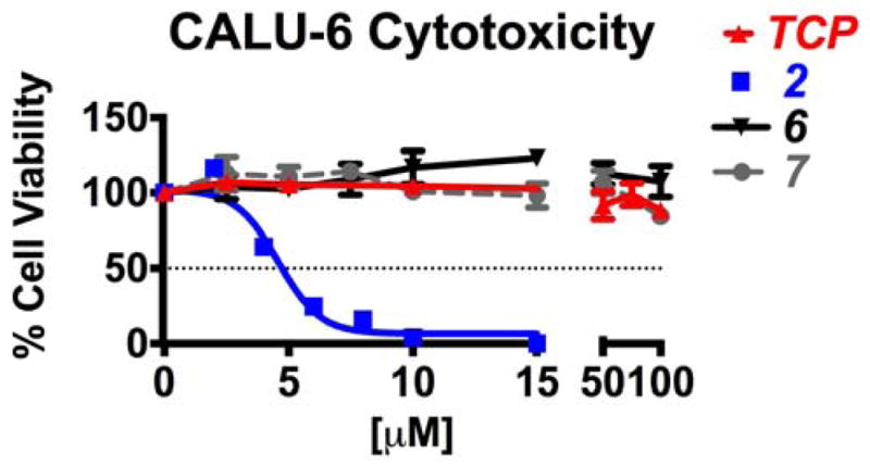
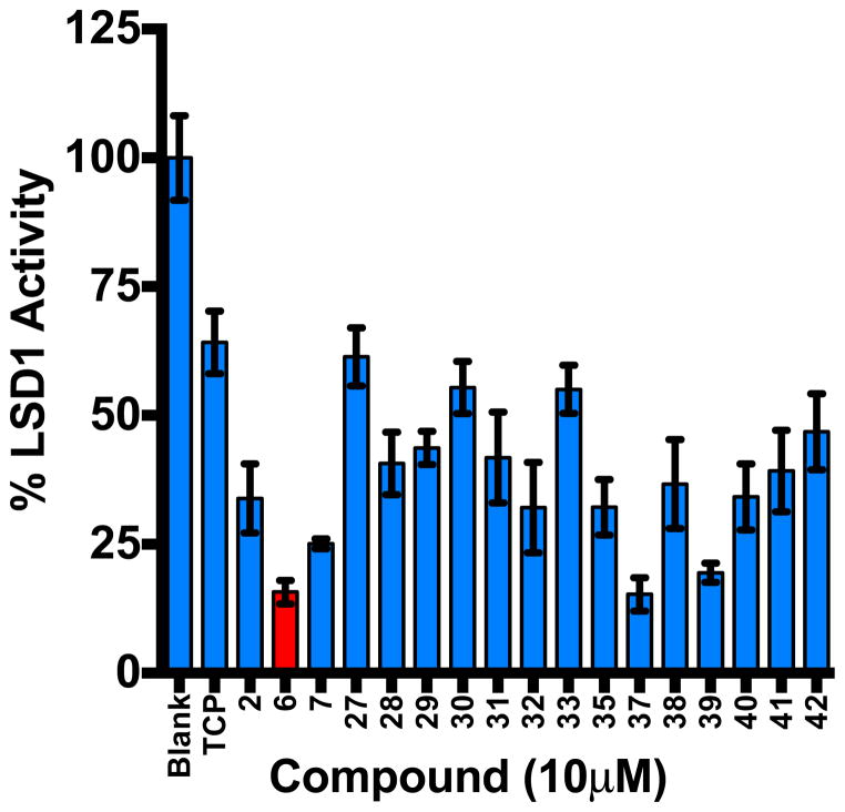
The chromatin remodeling amine oxidase lysine-specific demethylase 1 (LSD1) has become an attractive target for the design of specific inhibitors with therapeutic potential. We, and others, have described LSD1 inhibitors that have potential as antitumor agents. Many of the currently known LSD1 inhibitors are poor drug candidates, or are structurally based on the tranylcypromine backbone, thus increasing the potential for off-target effects mediated by other amine oxidases. We now describe a series of potent LSD1 inhibitors based on a novel 1,2,4-triazole scaffold; these inhibitors show a high degree of specificity for LSD1 in vitro, and cause increases in cellular histone 3 dimethyllysine 4 (H3K4me2), a gene transcription activating mark. Importantly, these inhibitors are not toxic to mammalian cells in vitro, and thus they may show utility in the treatment of epigenetically-based diseases where cell death is not a desired endpoint Figure 1. Structures of LSD1 inhibitors 1, verlindamycin 2, (bis)thioureas 3, amidoxime 4, cyclic peptide 5, N3-(2-chloro-6-phenoxybenzyl)-4H-1,2,4-triazole-3,5-diamine 6 and N3,N5-bis(2-methoxybenzyl)-1H-1,2,4-triazole-3,5-diamine 7.


Figures

Figure 1
Figure 1
Structures of LSD1 inhibitors 1, verlindamycin 2, (bis)thioureas 3, amidoxime 4, cyclic peptide 5, N3-(2-chloro-6-phenoxybenzyl)-4H-1,2,4-triazole-3,5-diamine 6 and N3, N-bis(2-methoxybenzyl)-1H-1,2,4-triazole-3,5-diamine 7.
Figure 2
Figure 2. Potency, inhibition kinetics, binding and selectivity of 3,5-diaminotriazoles 6 and 7
Panel A. Dose-response of LSD1 following treatment with compounds 6 and 7. Compound 6 IC50 = 1.19 μM; compound 7 IC50 = 2.22 μM Panel B. Competitive enzyme inhibition kinetics for LSD1 treated with compound 6 at 0, 0.375, 0.75, and 1 μM concentrations over a range of substrate concentrations between 0 and 100 μM. Ki for 6 = 2.2 μM Panel C. Isothermal calorimetry trace for LSD1 showing a release of heat upon titration with compound 6. Ka = 48.89 nM, R2=0.96 Panel D. MAO A and MAO B enzyme activity for 3,5-diaminotriazoles 6 and 7 compared to the known MAO inhibitor tranylcypromine (TCP). TCP IC50= 4.2 μM and 5.8 μM for MAO A and MAO B, respectively). Compounds 6 and 7 exhibited IC50 values > 100 μM for both MAO A and B. For Panels A. B and D, each data point is the average of 3 determination that in each case differed by 3% or less.
Figure 3
Figure 3
In silico analysis of compound 6 in the LSD1/CoREST catalytic site (PDB file 3ZMT). The aromatic portion of the o-phenoxy substituent lies 2.98Å from the FAD cofactor.
Figure 4
Figure 4. Cell viability and compound cytotoxicity in Calu-6
Cells were seeded at 3×10³ cells/well and treated 48–72 hrs with increasing concentrations of TCP, 2, 6 or 7. Cell viability was determined by colormetric CellTiter 96 Aqueous MTS (Promega, #G3580).
Figure 5
Figure 5. Global methylation changes by immunofluorescent staining
Calu-6 human lung adenocarcinoma cells were plated at 1×10³ cells/well and treated with 30μM TCP, 1- or 10μM compound 4, and 1- or 10μM compound 5 for 48hrs. Cells were stained for nucleus (DAPI), F-actin (Alexa Fluor 594 Phalloidin), and dimethyl-H3K4 (Alexa Fluor 488 Secondary Antibody). Fluorescent intensity on cell-by-cell basis was obtained by Hermes WiScan (IDEA Biomedical) and graphed as a frequency distribution histogram of relative cell count at specific intensities. Two-way ANOVA; *** P-value < 0.001, representative of 3 experiments with n=4–6 wells each.
Figure 6
Figure 6. LSD1 inhibition assay
Percent LSD1 activity remaining after treatment with 3,5-diaminotriazoles 6, 7, 2733, 35, 37–42 at 10 μM. Each data point is the average of 3 determinations ± SEM (see Table 2).
Scheme 1
Scheme 1

References

    1. Andreoli F, Del Rio A. Drug Disc Today. 2014 doi: 10.1016/j.drudis.2014.05.005. Epub ahead of print. - DOI - PubMed
    1. Shi Y, Lan F, Matson C, Mulligan P, Whetstine JR, Cole PA, Casero RA, Shi Y. Cell. 2004;119:941. - PubMed
    1. Suzuki T, Miyata N. J Med Chem. 2011;54:8236. - PubMed
    1. Varier RA, Timmers HT. Biochim Biophys Acta. 2011;1815:75. - PubMed
    1. Hayami S, Kelly JD, Cho HS, Yoshimatsu M, Unoki M, Tsunoda T, Field HI, Neal DE, Yamaue H, Ponder BA, Nakamura Y, Hamamoto R. Int’l J Cancer. 2011;128:574. - PubMed