. 2014 Mar 18:3:74.
doi: 10.12688/f1000research.3718.2. eCollection 2014.

Role of bacteriophages in STEC infections: new implications for the design of prophylactic and treatment approaches

Affiliations

Role of bacteriophages in STEC infections: new implications for the design of prophylactic and treatment approaches

Jaime H Amorim et al. F1000Res. .

Abstract

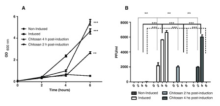
Shiga toxin (Stx) is considered the main virulence factor in Shiga toxin-producing Escherichia coli (STEC) infections. Previously we reported the expression of biologically active Stx by eukaryotic cells in vitro and in vivo following transfection with plasmids encoding Stx under control of the native bacterial promoter (1,2). Since stx genes are present in the genome of lysogenic bacteriophages, here we evaluated the relevance of bacteriophages during STEC infection. We used the non-pathogenic E. coli C600 strain carrying a lysogenic 933W mutant bacteriophage in which the stx operon was replaced by a gene encoding the green fluorescent protein (GFP). Tracking GFP expression using an In Vivo Imaging System (IVIS), we detected fluorescence in liver, kidney, and intestine of mice infected with the recombinant E. coli strain after treatment with ciprofloxacin, which induces the lytic replication and release of bacteriophages. In addition, we showed that chitosan, a linear polysaccharide composed of d-glucosamine residues and with a number of commercial and biomedical uses, had strong anti-bacteriophage effects, as demonstrated at in vitro and in vivo conditions. These findings bring promising perspectives for the prevention and treatment of haemolytic uremic syndrome (HUS) cases.


Conflict of interest statement

Competing interests: No competing interests were disclosed.

Figures

Figure 1.
Figure 1.. Induction of ϕΔTOX:GFP by ciprofloxacin and effect of chitosan in vitro.
A. Growth curve: the E. coli C600ΔTOX:GFP strain was induced with ciprofloxacin and the optical density was measured at 600nm at 0, 2, 4 and 6 hours after induction. Non-induced culture of E. coli C600ΔTOX:GFP strain was used as control. Chitosan was added at 2 or 4 hours after induction. B. Bacteriophage ϕΔTOX:GFP titers: bacteriophage titers were determined at 0, 2, 4 and 6 hours post-induction. Chitosan was added at 2 or 4 h post-induction. * p<0.05.
Figure 2.
Figure 2.. Detection of ϕΔTOX:GFP DNA in transfected mammalian cells.
A. PCR on DNA extracted from BHK-21 cells: 24 hours after transduction, BHK-21 cells were washed and treated with Trypsin-EDTA solution. DNA was extracted and PCR was performed. Lane 1: Cells transfected with ϕΔTOX:GFP. Lane 2: Cells transfected with ϕΔTOX:GFP previously treated with chitosan. Lane 3: Cells transfected with ϕΔTOX:GFP previously treated with DNAse. Lane 4. Untreated cells. Lane 5. Positive PCR control (ϕΔTOX:GFP DNA). Lane 6. Negative PCR control. Lane 7. 1 kb ladder (Invitrogen).
Figure 3.
Figure 3.. Detection of in vivo GFP expression in mice infected with the lysogenic E. coli C600ΔTOX:GFP strain using In Vivo Imaging System (IVIS).
A. IVIS Representative image: ciprofloxacin was administered 2 hours post-infection to induce ϕΔTOX:GFP in vivo. Mice were treated with chitosan 2 hours after bacteriophage induction. All mice were sacrificed 24 hours post-infection and brains, hearts, lungs, livers, spleens, kidneys and intestines were harvested and analyzed by IVIS. Fluorescence intensity was recorded as photons/sec/cm 2, and the signal intensity represents the amount of GFP present. B. Graphic of fluorescence intensity on GFP-positive organs. Four animals per group were analyzed and the fluorescence intensity was quantified using Living Imaging 4.3.1 in Calipter Life Sciences.
Figure 4.
Figure 4.. Treatment with chitosan delays death of mice infected with the EHEC EDL933W strain.
Mice were infected orally with the EHEC EDL933W strain. Controls did not receive chitosan (dots and broken line) and the experimental group received chitosan 2 hours post-infection (square and fill line). Survival rates were followed for one week.

References

    1. Bentancor LV, Bilen MF, Mejías MP, et al. : Functional capacity of Shiga-toxin promoter sequences in eukaryotic cells. PLoS One. 2013;8(2):e57128. 10.1371/journal.pone.0057128 - DOI - PMC - PubMed
    1. Bentancor LV, Mejías MP, Pinto A, et al. : Promoter sequence of Shiga toxin 2 (Stx2) is recognized in vivo leading to production of biologically active Stx2. MBio. 2013;4(5):e00501–13. 10.1128/mBio.00501-13 - DOI - PMC - PubMed
    1. Petruzziello-Pellegrini TN, Marsden PA: Shiga toxin-associated hemolytic uremic syndrome: advances in pathogenesis and therapeutics. Curr Opin Nephrol Hypertens. 2012;21(4):433–40. 10.1097/MNH.0b013e328354a62e - DOI - PMC - PubMed
    1. Tyler JS, Beeri K, Reynolds JL, et al. : Prophage induction is enhanced and required for renal disease and lethality in an EHEC mouse model. PLoS Pathog. 2013;9(3):e1003236. 10.1371/journal.ppat.1003236 - DOI - PMC - PubMed
    1. Kong M, Chen XG, Xing K, et al. : Antimicrobial properties of chitosan and mode of action: a state of the art review. Int J Food Microbiol. 2010;144(1):51–63. 10.1016/j.ijfoodmicro.2010.09.012 - DOI - PubMed

LinkOut - more resources