. 2014:2014:732096.
doi: 10.1155/2014/732096. Epub 2014 Dec 11.

Osteoma of the pharynx that developed from the hyoid bone

Affiliations

Osteoma of the pharynx that developed from the hyoid bone

Akira Hagiwara et al. Case Rep Otolaryngol. 2014.

Abstract

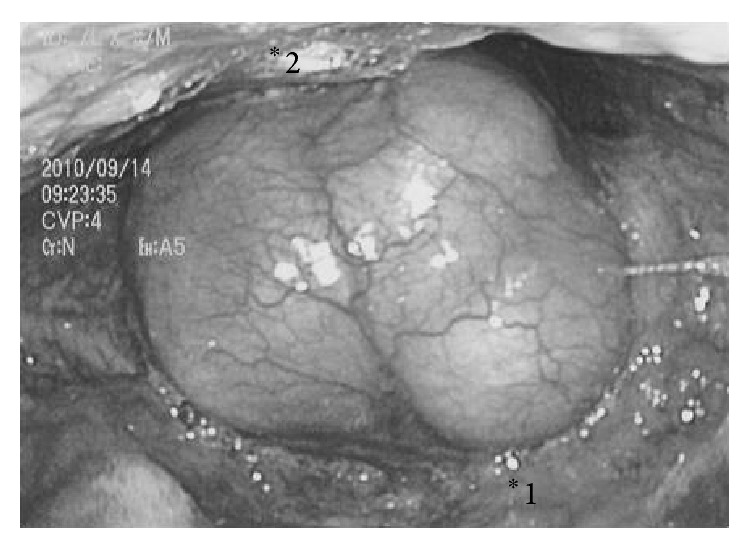
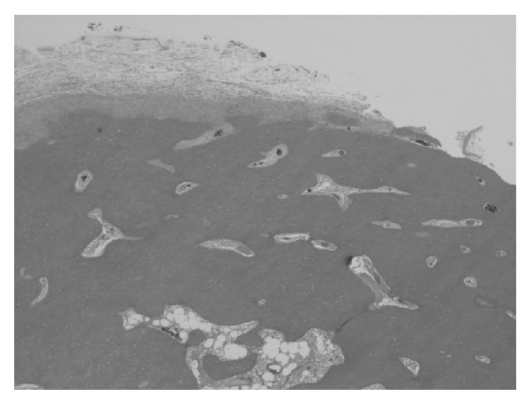
This paper reports on apparently the first case of a pharyngeal osteoma that developed from the hyoid bone. An 84-year-old man's, presenting symptom was a slight throat pain. Endoscopic examination revealed a huge mass occluding the pharyngeal space. CT scan of the neck showed a large osseous mass adjacent to the hyoid bone. Transoral resection with tracheostomy was performed. Histopathologically, the tumor consisted of mature lamellar bone without a fibrous component. For two years postoperatively, the patient has been free from throat symptoms and signs of recurrence. Osteomas are benign, slow-growing tumors. They rarely develop symptoms or cause functional disturbance. We performed total resection to avoid further functional disturbance as the osteoma was huge. To the best of our knowledge, this is the first report on an osteoma that occupied the pharyngeal space and developed from the hyoid bone.


Figures

Figure 1
Figure 1
Endoscopic examination of the pharynx. *1: root of the tongue; *2: posterior wall of the pharynx.
Figure 2
Figure 2
CT scan of the neck. A large osseous mass adjacent to the hyoid bone was observed.
Figure 3
Figure 3
Tumor size and diameter. The tumor was round and 4 cm in diameter. An irregular osseous mass covered with mucosa was observed.
Figure 4
Figure 4
Histological examination of the tumor (H&E ×20). The tumor consisted of mature lamellar bone without a fibrous component. Bone marrow cells and trabeculae were confirmed in the center of the tumor.

References

    1. Woldenberg Y., Nash M., Bodner L. Peripheral osteoma of the maxillofacial region. Diagnosis and management: a study of 14 cases. Medicina Oral, Patología Oral y Cirugía Bucal. 2005;10:E139–142. - PubMed
    1. Horikawa F. K., de Freitas R. R., Maciel F. A., Gonçalves A. J. Peripheral osteoma of the maxillofacial region: a study of 10 cases. Brazilian Journal of Otorhinolaryngology. 2012;78(5):38–43. doi: 10.5935/1808-8694.20120006. - DOI - PMC - PubMed
    1. Angelillo M., Mazzone S., Costa G., Mazzone A., Barillari U. The first case of osteoma in the false vocal fold. Auris Nasus Larynx. 2009;36(2):235–238. doi: 10.1016/j.anl.2008.04.014. - DOI - PMC - PubMed
    1. Ben-Yaakov A., Wohlgelernter J., Gross M. Osteoma of the lateral semicircular canal. Acta Oto-Laryngologica. 2006;126(9):1005–1007. doi: 10.1080/00016480500527292. - DOI - PubMed
    1. Jang C. H., Cho Y. B. Osteoma of the incus with congenital cholesteatoma: a case report. Auris Nasus Larynx. 2009;36(3):349–352. doi: 10.1016/j.anl.2008.08.002. - DOI - PubMed

LinkOut - more resources