. 2014:5:1000256.
doi: 10.4172/2155-9899.1000256.

Corneal Lymphatics: Role in Ocular Inflammation as Inducer and Responder of Adaptive Immunity

Affiliations

Corneal Lymphatics: Role in Ocular Inflammation as Inducer and Responder of Adaptive Immunity

Sunil K Chauhan et al. J Clin Cell Immunol. 2014.

Abstract

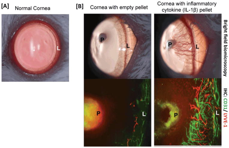
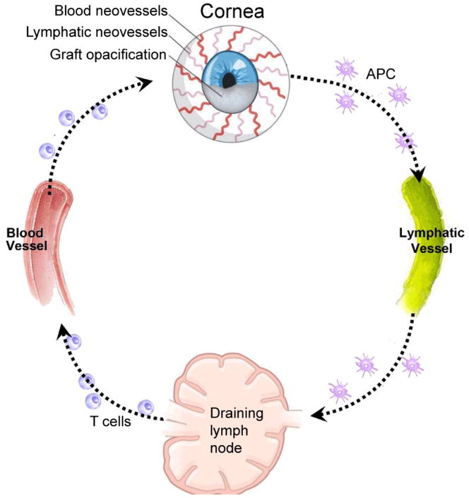
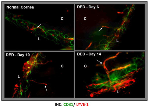
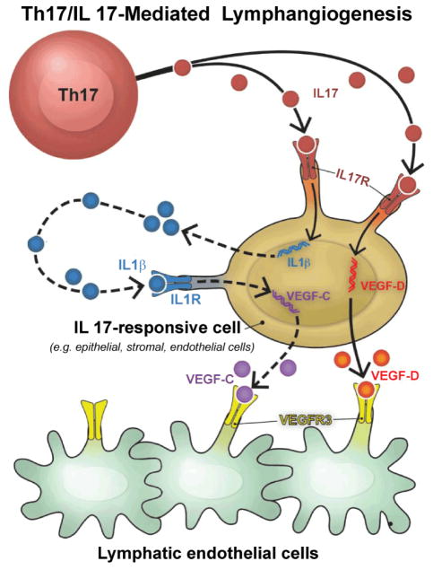
The normal cornea is devoid of lymphatic and blood vessels, thus suppressing both the afferent (lymphatic) and efferent (vascular) arms of the immune response-contributing to its 'immune privilege'. Inflammation, however, negates this unique 'immune' and 'angiogenic' privilege of the cornea. Abnormal blood vessel growth from pre-existing limbal vessels into the cornea has been studied for many years, but it is only recently that the significance of new lymphatic vessels (lymphangiogenesis) in ocular inflammatory diseases has been demonstrated. Whereas blood vessels in inflamed ocular surface provide a route of entry for immune effector cells to the cornea, lymphatics facilitate the exit of antigen-presenting cells and antigenic material from the cornea to regional lymph nodes, thus promoting induction of adaptive immune response. This review summarizes the current evidence for lymphangiogenesis in the cornea, and describes its molecular mediators; and discusses the interface between corneal lymphangiogenesis and adaptive immunity. Furthermore, the pathophysiologic implications of corneal lymphangiogenesis in the setting of allo- and autoimmune-mediated corneal inflammation are discussed.

Keywords: Adaptive immunity; Cornea; Lymphangiogenesis; Ocular inflammation.


Figures

Figure 1
Figure 1
Angiogenic privilege of cornea. [A] Normal cornea is a blood and lymphatic vessel-free tissue. Vessels are restricted only to the periphery of the cornea (limbal area). [B] In response to inflammation, this “angiogenic” privilege of the cornea can be lost leading to the ingrowth of both blood and lymphatic vessels. Micrographs from a mouse corneal micropocket model of neovascularization show that pellets containing the inflammatory cytokine IL-1β robustly induce ingrowth of both new blood (CD31+LYVE1) and lymphatic (LYVE1+CD31lo) vessels. (L: Limbus; P: Pellet).
Figure 2
Figure 2
Function of lymphatic and blood vessels in corneal alloimmunity. Lymphatics (afferent arm of immune system) transport antigens and antigen-presenting cells (APCs) from the graft site to the draining lymph nodes, where T cells are primed and expanded. Alloreactive T cells return to the cornea via blood vessels (efferent arm) and mediate transplant rejection.
Figure 3
Figure 3
Selective ingrowth of corneal lymphatic vessels in dry eye disease. Confocal micrographs showing corneal lymphangiogenesis in normal cornea and in dry eye corneas at days 6, 10, and 14 post-induction of dry eye in a mouse model of dry eye disease. The lymphatic vessels (LYVE1+CD31lo) increase both in area and caliber, and grow towards the cornea center with disease progression. The lymphatics are unaccompanied by blood vessels (CD31hi/LYVE1). (Lymphatics marked by arrows; C: Cornea; L: Limbus). Adapted from [81].
Figure 4
Figure 4
Mechanism of T-helper-17 (Th17) cell-mediated lymphangiogenesis. Th17 cell-secreted interleukin-17 (IL-17) induces lymphangiogenesis via a vascular endothelial growth factor (VEGF)-D/C–VEGFR-3 signaling pathway. In response to Th17-secreted IL-17, IL-17-receptor-expressing cells such as epithelial and stromal cells directly upregulate expression of VEGF-D; whereas VEGF-C expression is upregulated via IL-17-induced IL-1β-mediated pathway. Both VEGF-D and VEGF-C bind to VEGFR-3 on lymphatic endothelial cells and promote proliferation and tube formation by lymphatic endothelial cells.

References

    1. Lee P, Wang CC, Adamis AP. Ocular neovascularization: an epidemiologic review. Surv Ophthalmol. 1998;43:245–269. - PubMed
    1. Hos D, Bachmann B, Bock F, Onderka J, Cursiefen C. Age-related changes in murine limbal lymphatic vessels and corneal lymphangiogenesis. Exp Eye Res. 2008;87:427–432. - PubMed
    1. Cursiefen C, Chen L, Borges LP, Jackson D, Cao J, et al. VEGF-A stimulates lymphangiogenesis and hemangiogenesis in inflammatory neovascularization via macrophage recruitment. J Clin Invest. 2004;113:1040–1050. - PMC - PubMed
    1. Lohela M, Bry M, Tammela T, Alitalo K. VEGFs and receptors involved in angiogenesis versus lymphangiogenesis. Curr Opin Cell Biol. 2009;21:154–165. - PubMed
    1. Mäkinen T, Veikkola T, Mustjoki S, Karpanen T, Catimel B, et al. Isolated lymphatic endothelial cells transduce growth, survival and migratory signals via the VEGF-C/D receptor VEGFR-3. EMBO J. 2001;20:4762–4773. - PMC - PubMed

LinkOut - more resources